. 2019 Jul 17:13:70.
doi: 10.3389/fnana.2019.00070. eCollection 2019.

Brain Organization of Apolygus lucorum: A Hemipteran Species With Prominent Antennal Lobes

Affiliations

Brain Organization of Apolygus lucorum: A Hemipteran Species With Prominent Antennal Lobes

Gui-Ying Xie et al. Front Neuroanat. .

Abstract

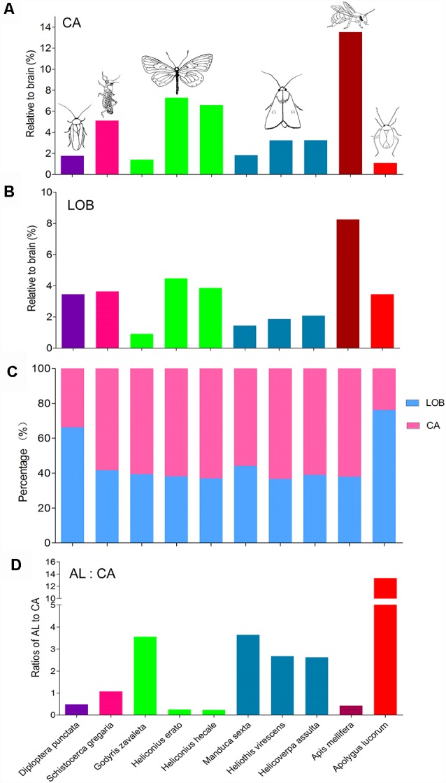
The anatomical organization of distinct regions in the insect brain often reflects their functions. In the present study, the brain structure of Apolygus lucorum was examined by using immunolabeling and three-dimensional reconstruction. The results revealed the location and volume of prominent neuropils, such as the antennal lobes (AL), optic lobes (OL), anterior optic tubercles (AOTU), central body (CB), lateral accessory lobes (LAL), mushroom lobes, and distinct tritocerebral neuropils. As expected, this brain is similar to that of other insects. One exception, however, is that the antennal lobes were found to be the most prominent neuropils. Their size relative to the entire brain is the largest among all insect species studied so far. In contrast, the calyx, a region getting direct input from the antennal lobe, has a smaller size relative to the brain than that of other species. These findings may suggest that olfaction plays an essential role for A. lucorum.

Keywords: Apolygus lucorum; anatomy; antennal lobe; brain; neuropil.


Figures

Figure 1
Figure 1
Three-dimensional reconstructions and confocal images of the Apolygus lucorum brain. (A) Three-dimensional reconstructions of brain in frontal view. (B) Posterior view. (C) Lateral view. (D–G) Serial confocal images showing the brain sections of A. lucorum. AMMC, antennal mechanosensory and motor center; AL, antennal lobe; AOTU, anterior optic tubercle; CA, calyx; CB, central body; ES, esophagus; GNG, gnathal ganglion; LA, lamina; LOB, mushroom body lobes; LOX, lobula complex; ME, medulla; PB, protocerebral bridge; PED, pedunculus; TR, tritocerebrum. Directions: a, anterior; d, dorsal; l, lateral; m, medial; p, posterior, v, ventral. Scale bars: 100 μm.
Figure 2
Figure 2
Three-dimensional reconstructions and confocal images of the Apolygus lucorum antennal lobe and antennal lobe tracts. (A) Three-dimensional reconstructions of antennal lobe glomerulus in dorsal view. (B) Vental view. (C) Lateral view. (D,E) Confocal images of antennal lobe glomeruli at different depths. (F,G) Three-dimensional reconstructions of antennal lobe tracts in dorsal view and ventral respectively. (H–J) Confocal images of antennal lobe tracts and targeted regions at different depths. AL, antennal lobe; CA, calyx; CB, central body; ES, esophagus; GNG, gnathal ganglion; lALT, lateral antennal lobe tract; LH, lateral horn; mALT, medial antennal lobe tract; mlALT, medial lateral antennal lobe tract; sALT, small antennal lobe tract. Directions: a, anterior; d, dorsal; l, lateral; m, medial; p, posterior, v, ventral. Scale bars: 50 μm.
Figure 3
Figure 3
Three-dimensional reconstructions and confocal images of Apolygus lucorum optic lobe and optic tubercles. (A) Three-dimensional reconstructions of optic lobe in frontal view. (B) Posterior view. (C) Sagittal view. (D–F) Confocal images of optic lobe at different depths. (G) Three-dimensional reconstructions of anterior and posterior optic tubercles in frontal view. (H) Posterior view. (I) Dorsal view. (J,K) Confocal images of AOTU in dorsal view. (L,M) Confocal images of AOTU in frontal view. (N) A confocal image of posterior optic tubercle in frontal view. (O) A confocal image of posterior optic tubercle in dorsal view. AME, accessory medulla; AOT, anterior optic tract; DLO-A, anterior part of dorsal lobula; DLO-P, posterior part of dorsal lobula; ES, esophagus; LA, lamina; LU, lower unit of anterior optic tubercle; ME, medulla; MLO, medial lobula; PB, protocerebral bridge; POTU, posterior optic tubercle; PS, posterior slope; UU, upper unit of anterior optic tubercle; VLO, ventral lobula. Directions: a, anterior; d, dorsal; l, lateral; m, medial; p, posterior; v, ventral. Scale bars: 100 μm in (A–C,G–I,L–O), 50 μm in (D–F,J,K).
Figure 4
Figure 4
Three-dimensional reconstructions and confocal images of Apolygus lucorum CB, PB, and LAL. (A) Three-dimensional reconstructions of CB, PB, and LAL in dorsal view. (B) Anterior view. (C) Three-dimensional reconstructions of PB and calyces in posterior view. (D) A confocal image showing the CB. (E) A confocal image showing the CB and LAL. (F) A confocal image showing the PB and calyces. AL, antennal lobe; CA, calyx; CBL, lower unit of central body; CBU, upper unit of central body; LAL, lateral accessory lobe; OL, optic lobe; PB, protocerebrum bridge. Directions: a, anterior; d, dorsal; l, lateral; p, posterior; v, ventral. Scale bars: 100 μm in (A,B,D,E), 50 μm in (C,F).
Figure 5
Figure 5
Three-dimensional reconstructions and confocal images of Apolygus lucorum mushroom body. (A) Three-dimensional reconstructions of mushroom body in dorsal view. (B) Three-dimensional reconstruction of calyx in anterior view. (C) A confocal image showing the calyx. (D) Three-dimensional reconstructions of mushroom body in dorsal view. (E) Frontal view. (F) ventral view. (G–J) Confocal images at different depths showing mushroom body lobes of one preparation. (K–N) Confocal images at different depths showing mushroom body lobes of the other preparation. AL, antennal lobe; CA, calyx; CAL, lateral calyx; CAM, medial calyx; CB, central body; PED, pedunculus; PEDL, lateral pedunculus; PEDM, medial pedunculus; α, α lobe; β, β lobe; βa–βe, a–e five β lobelets; γ, γ lobe; γa and γb, a and b two γ lobelets. Scale bars: 100 μm in (A), 50 μm in (B–N).
Figure 6
Figure 6
Absolute volumes and relative sizes of prominent neuropils in the brain of Apolygus lucorum. (A) Absolute volumes. (B) Relative size (i.e., size relative to the volume of the whole brain). AL, antennal lobe; AOTU, anterior optic tubercle; CA, calyx; CB, central body; GNG, gnathal ganglion; LA, lamina; LAL, lateral accessory lobe; LOB, mushroom body lobes; LOX, lobula complex; ME, medulla; PB, protocerebral bridge; PED, pedunculus; TR, tritocereburm. Midbrain, the remaining neuropils in the central brain except MB, CB, PB, LAL, and AOTU.
Figure 7
Figure 7
Comparison of relative sizes of the antennal lobe, optic lobe, CB, and mushroom body to the whole brain in different species. (A) Antennal lobe. (B) Optic lobe including medulla and lobula complex. (C) CB. (D) Mushroom body. AL, antennal lobe; CB, central body; MB, mushroom body; OL, optic lobe. *Data missing.
Figure 8
Figure 8
Comparison of relative sizes of the calyx and lobes in different species. (A) Relative sizes of the calyx to the whole brain in different species. (B) Relative sizes of the mushroom body lobes to the whole brain in different species. (C) Proportion of calyx and lobes to the whole mushroom body. (D) The ratios of antennal lobe to calyx in different species. AL, antennal lobe; CA, calyx; LOB, mushroom body lobes including the pedunculus.

References

    1. Anton S., Homberg U. (1999). “Antennal lobe structure,” in Insect Olfaction, ed. Hansson B. S. (Berlin: Springer; ), 97–124.
    1. Barrozo R., Couton L., Lazzari C. R., Insausti T., Minoli S. A., Fresquet N., et al. . (2009). Antennal pathways in the central nervous system of a blook-sucking bug, Rhodnius prolixus. Arthropod Struct. Dev. 38, 101–110. 10.1016/j.asd.2008.08.004 - DOI - PubMed
    1. Beetz M. J., el Jundi B., Heinze S., Homberg U. (2015). Topographic organization and possible function of the posterior optic tubercles in the brain of the desert locust Schistocerca gregaria. J. Comp. Neurol. 523, 1589–1607. 10.1002/cne.23736.2018 - DOI - PubMed
    1. Brandt R., Rohlfing T., Rybak J., Krofczik S., Maye A., Westerhoff M., et al. . (2005). Three-dimensional average-shape atlas of the honeybee brain and its applications. J. Comp. Neurol. 492, 1–19. 10.1002/cne.20644 - DOI - PubMed
    1. Bressan J. M., Benz M., Oettler J., Heinze J. J., Hartenstein V., Sprecher S. G. (2015). A map of brain neuropils and fiber systems in the ant Cardiocondyla obscurior. Front. Neuroanat. 8:166. 10.3389/fnana.2014.00166 - DOI - PMC - PubMed