. 2019 Jul 16:10:726.
doi: 10.3389/fneur.2019.00726. eCollection 2019.

Measurement Variability Following MRI System Upgrade

Affiliations

Measurement Variability Following MRI System Upgrade

Olivier Potvin et al. Front Neurol. .

Abstract

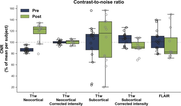
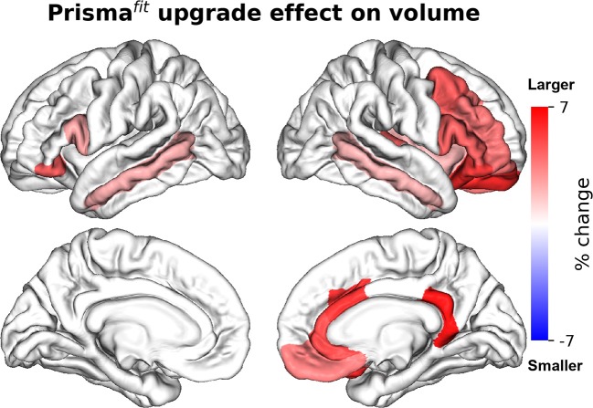
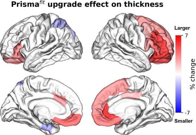
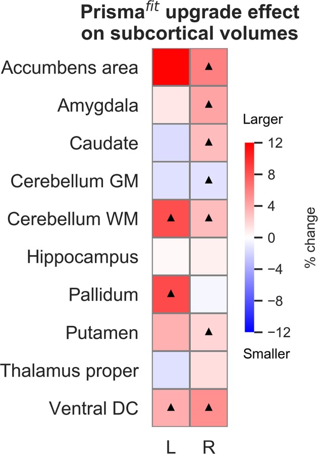
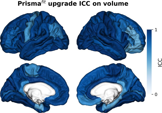
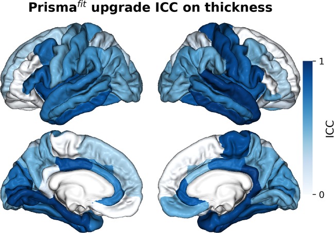
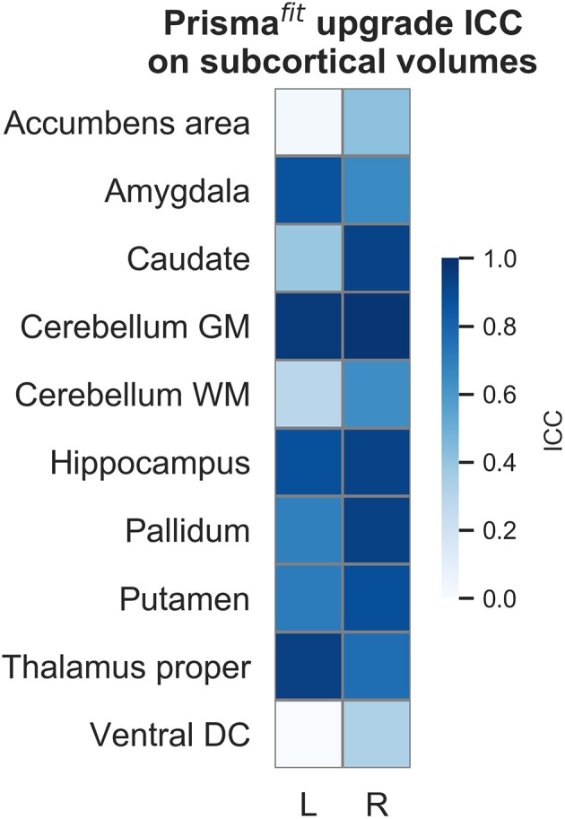
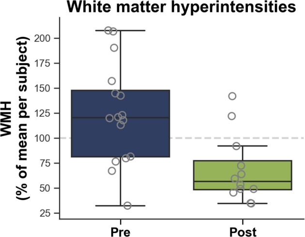
Major hardware/software changes to MRI platforms, either planned or unplanned, will almost invariably occur in longitudinal studies. Our objective was to assess the resulting variability on relevant imaging measurements in such context, specifically for three Siemens Healthcare Magnetom Trio upgrades to the Prismafit platform. We report data acquired on three healthy volunteers scanned before and after three different platform upgrades. We assessed differences in image signal [contrast-to-noise ratio (CNR)] on T1-weighted images (T1w) and fluid-attenuated inversion recovery images (FLAIR); brain morphometry on T1w image; and small vessel disease (white matter hyperintensities; WMH) on FLAIR image. Prismafit upgrade resulted in higher (30%) and more variable neocortical CNR and larger brain volume and thickness mainly in frontal areas. A significant relationship was observed between neocortical CNR and neocortical volume. For FLAIR images, no significant CNR difference was observed, but WMH volumes were significantly smaller (-68%) after Prismafit upgrade, when compared to results on the Magnetom Trio. Together, these results indicate that Prismafit upgrade significantly influenced image signal, brain morphometry measures and small vessel diseases measures and that these effects need to be taken into account when analyzing results from any longitudinal study undergoing similar changes.

Keywords: MRI upgrade; Siemens healthcare; longitudinal studies; magnetic resonance imaging; morphometry; neuroimaging; variability.


Figures

Figure 1
Figure 1
Contrast-to-noise ratio (CNR) before (Pre) and after (Post) Prismafit upgrade from T1w images and FLAIR images.
Figure 2
Figure 2
Percentage of change in neocortical regions' volume after Prismafit upgrade. Only regions that were significant after false-discovery rate correction (p < 0.05) are showed.
Figure 3
Figure 3
Percentage of change in neocortical regions' thickness after Prismafit upgrade. Only regions that were significant after false-discovery rate correction (p < 0.05) are showed.
Figure 4
Figure 4
Percentage of change in subcortical regions' volume after Prismafit upgrade. Black triangles denote regions that were significant after false-discovery rate correction (p < 0.05).
Figure 5
Figure 5
Intraclass correlations (ICC) of neocortical regions' volume before and after Prismafit upgrade.
Figure 6
Figure 6
Intraclass correlations (ICC) of neocortical regions' thickness before and after Prismafit upgrade.
Figure 7
Figure 7
Intraclass correlations (ICC) of subcortical regions' volume before and after Prismafit upgrade.
Figure 8
Figure 8
White matter hyperintensities (WMH) measured on FLAIR images according to before (Pre) and after (Post) Prismafit upgrade.

References

    1. Potvin O, Dieumegarde L, Duchesne S. Normative morphometric data for cerebral cortical areas over the lifetime of the adult human brain. Neuroimage. (2017) 156:315–39. 10.1016/j.neuroimage.2017.05.019 - DOI - PubMed
    1. Potvin O, Dieumegarde L, Duchesne S. Freesurfer cortical normative data for adults using Desikan-Killiany-Tourville and ex vivo protocols. Neuroimage. (2017) 156:43–64. 10.1016/j.neuroimage.2017.04.035 - DOI - PubMed
    1. Govindarajan KA, Freeman L, Cai C, Rahbar MH, Narayana PA. Effect of intrinsic and extrinsic factors on global and regional cortical thickness. PLoS ONE. (2014) 9:e96429. 10.1371/journal.pone.0096429 - DOI - PMC - PubMed
    1. Han X, Jovicich J, Salat D, van der Kouwe A, Quinn B, Czanner S, et al. . Reliability of MRI-derived measurements of human cerebral cortical thickness: the effects of field strength, scanner upgrade and manufacturer. Neuroimage. (2006) 32:180–94. 10.1016/j.neuroimage.2006.02.051 - DOI - PubMed
    1. Kruggel F, Turner J, Muftuler LT. Impact of scanner hardware and imaging protocol on image quality and compartment volume precision in the ADNI cohort. Neuroimage. (2010) 49:2123–33. 10.1016/j.neuroimage.2009.11.006 - DOI - PMC - PubMed

LinkOut - more resources