

Interaction Between Staphylococcal Biofilm and Bone: How Does the Presence of Biofilm Promote Prosthesis Loosening?
- PMID: 31379772
- PMCID: PMC6653651
- DOI: 10.3389/fmicb.2019.01602
Interaction Between Staphylococcal Biofilm and Bone: How Does the Presence of Biofilm Promote Prosthesis Loosening?
Abstract
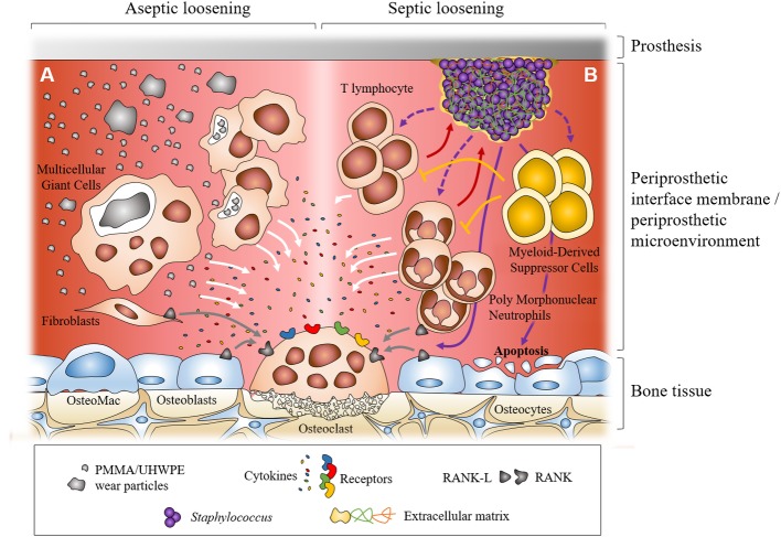
With the aging of population, the number of indications for total joint replacement is continuously increasing. However, prosthesis loosening can happen and is related to two major mechanisms: (1) aseptic loosening due to prosthesis micromotion and/or corrosion and release of wear particles from the different components of the implanted material and (2) septic loosening due to chronic prosthetic joint infection (PJI). The "aseptic" character of prosthesis loosening has been challenged over the years, especially considering that bacteria can persist in biofilms and be overlooked during diagnosis. Histological studies on periprosthetic tissue samples reported that macrophages are the principle cells associated with aseptic loosening due to wear debris. They produce cytokines and favor an inflammatory environment that induces formation and activation of osteoclasts, leading to bone resorption and periprosthetic osteolysis. In PJIs, the presence of infiltrates of polymorphonuclear neutrophils is a major criterion for histological diagnosis. Neutrophils are colocalized with osteoclasts and zones of osteolysis. A similar inflammatory environment also develops, leading to bone resorption through osteoclasts. Staphylococcus aureus, Staphylococcus epidermidis, and Staphylococcus lugdunensis are the main staphylococci observed in PJIs. They share the common feature to form biofilm. For S. aureus and S. epidermidis, the interaction between biofilm and immunes cells (macrophages and polymorphonuclear neutrophils) differs regarding the species. Indeed, the composition of extracellular matrix of biofilm seems to impact the interaction with immune cells. Recent papers also reported the major role of myeloid-derived suppressor cells in biofilm-associated PJIs with S. aureus. These cells prevent lymphocyte infiltration and facilitate biofilm persistence. Moreover, the role of T lymphocytes is still unclear and potentially underestimates. In this review, after introducing the cellular mechanism of aseptic and septic loosening, we will focus on the interrelationships between staphylococcal biofilm, immune cells, and bone cells.
Keywords: Staphylococcus; biofilm; osteoblast; osteoclast; polymorphonuclear neutrophil; prosthesis loosening; prosthetic joint infection.
Figures
References
-
- Albrektsson T., Albrektsson B. (1987). Osseointegration of bone implants. A review of an alternative mode of fixation. Acta Orthop. Scand. 58, 567–577. - PubMed
-
- Albrektsson T., Chrcanovic B., Jacobsson M., Wennerberg A. (2017). Osseointegration of implants – a biological and clinical overview. JSM Dent. Surg. 2:1022.
Publication types
LinkOut - more resources
Full Text Sources