. 2019 Jul 16:10:1631.
doi: 10.3389/fmicb.2019.01631. eCollection 2019.

Type III Secretion System of Beneficial Rhizobacteria Pseudomonas simiae WCS417 and Pseudomonas defensor WCS374

Affiliations

Type III Secretion System of Beneficial Rhizobacteria Pseudomonas simiae WCS417 and Pseudomonas defensor WCS374

Ioannis A Stringlis et al. Front Microbiol. .

Abstract

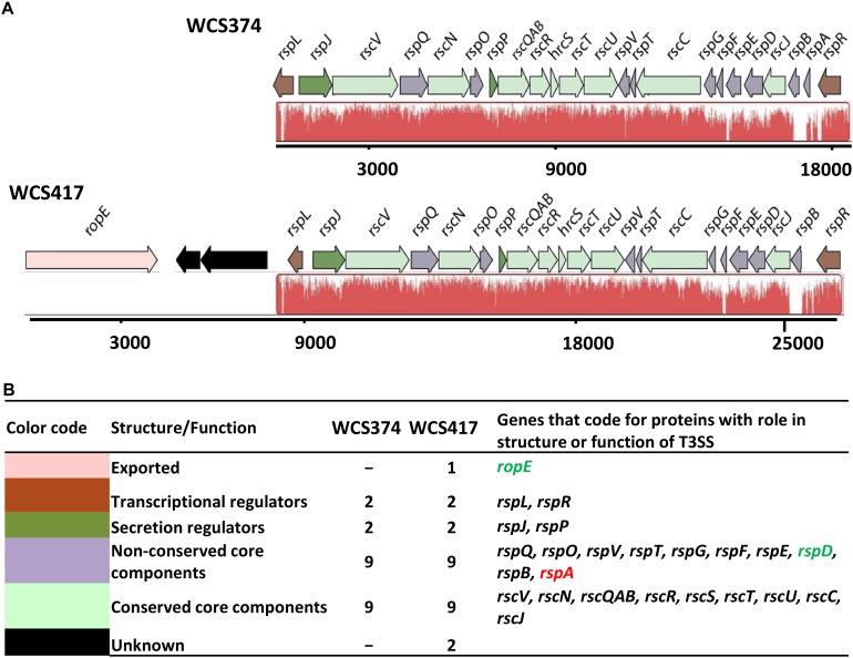
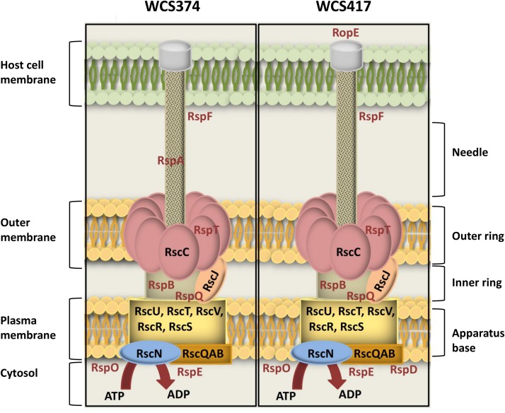
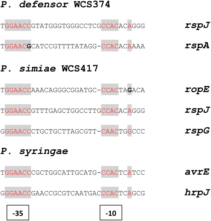
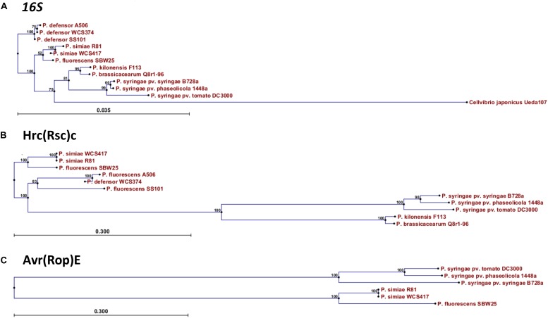
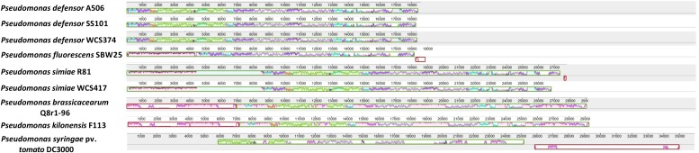
Plants roots host myriads of microbes, some of which enhance the defense potential of plants by activating a broad-spectrum immune response in leaves, known as induced systemic resistance (ISR). Nevertheless, establishment of this mutualistic interaction requires active suppression of local root immune responses to allow successful colonization. To facilitate host colonization, phytopathogenic bacteria secrete immune-suppressive effectors into host cells via the type III secretion system (T3SS). Previously, we searched the genomes of the ISR-inducing rhizobacteria Pseudomonas simiae WCS417 and Pseudomonas defensor WCS374 for the presence of a T3SS and identified the components for a T3SS in the genomes of WCS417 and WCS374. By performing a phylogenetic and gene cluster alignment analysis we show that the T3SS of WCS417 and WCS374 are grouped in a clade that is enriched for beneficial rhizobacteria. We also found sequences of putative novel effectors in their genomes, which may facilitate future research on the role of T3SS effectors in plant-beneficial microbe interactions in the rhizosphere.

Keywords: beneficial rhizobacteria; effectors; induced systemic resistance; rhizosphere; type III secretion system.


Figures

FIGURE 1
FIGURE 1
Genetic organization of the T3SS gene clusters of WCS347 and WCS417. (A) Orientation and organization of the genes in the T3SS clusters recognized in the genomic sequence of P. defensor WCS374 (WCS374) and P. simiae WCS417 (WCS417). The red color below the genes indicate regions similar between the two clusters, and the height of the peaks the degree of similarity. (B) Each gene in the cluster in panel (A) is given a color based on the role its encoding protein has either in the structure or the function of the T3SS apparatus. The color code, the putative role of the protein encoded by each gene, number of genes encoding for proteins with specific role and their names are presented. Green letters indicate gene presence only in the WCS417 cluster, while red letters indicate gene presence only in the WCS374 cluster. The numbers below the gene clusters are indicative of the length of the cluster.
FIGURE 2
FIGURE 2
Schematic overview of T3SS-injection machineries of WCS347 and WCS417. Conserved and non-conserved components of the T3SS-body were identified by BlastP and the encoded proteins are placed as parts of the T3SS injectisome, based on their P. syringae homologs (Galán et al., 2014). On the right of the figure the basic parts of T3SS body are indicated (apparatus base, inner ring, outer ring and needle). On the left, the cell compartments where the T3SS is spanning from including the bacterial cytosol, plasma membrane and outer membrane and the cell membrane of the host. Conserved components are indicated with black letters, and non-conserved with red letters.
FIGURE 3
FIGURE 3
Structure and position of putative hrp boxes identified in WCS374 and WCS417 gene clusters. Putative RspL- binding sites in genes present in the T3SS gene clusters of WCS417 and WCS374 found after manual examination in the promoter region upstream of the indicated genes. Conserved nucleotides are indicated with red letters and divergent nucleotides with bold letters. The box motif upstream of ropE, similarly to avrE, have a shorter spacer region of 15 nucleotides between the –35 and –10 sites.
FIGURE 4
FIGURE 4
Phylogenetic relationship of WCS417 and WCS374 T3SS components with other beneficial and phytopathogenic Pseudomonas spp. Neighbor-joining phylogenetic trees for aligned nucleotide sequence of 16S rRNA (A) and protein sequences of Hrc(Rsc)C (B) and Avr(Rop)E (C). In the case of 16S rRNA the evolutionary distance was calculated with Kimura 80 model, while evolutionary distance between protein sequences was estimated with Jukes-Cantor model. Numbers on nodes are calculated with bootstrap test using 1000 replicates. The length of branches is corresponding to the amount of changes in the time of evolution.
FIGURE 5
FIGURE 5
Similarity of WCS417 and WCS374 T3SS gene clusters with other beneficial and phytopathogenic Pseudomonas spp. Gene clusters were identified in available sequenced genomes of beneficial and phytopathogenic Pseudomonas spp. Progressive Mauve was used to align the clusters and find regions exhibiting high similarity. Similar colors correspond to similar genomic regions in a cluster. The height of peaks in each cluster depicts the degree of similarity in regions present in T3SS clusters of different Pseudomonas spp.

References

    1. Alfano J. R., Collmer A. (2004). Type III secretion system effector proteins: double agents in bacterial disease and plant defense. Annu. Rev. Phytopathol. 42 385–414. 10.1146/annurev.phyto.42.040103.110731 - DOI - PubMed
    1. Almario J., Bruto M., Vacheron J., Prigent-Combaret C., Moenne-Loccoz Y., Muller D. (2017). Distribution of 2,4-diacetylphloroglucinol biosynthetic genes among the Pseudomonas spp. Reveals unexpected polyphyletism. Front. Microbiol. 8:1218. 10.3389/fmicb.2017.01218 - DOI - PMC - PubMed
    1. Anderson J. C., Wan Y., Kim Y. M., Pasa-Tolic L., Metz T. O., Peck S. C. (2014). Decreased abundance of type III secretion system-inducing signals in Arabidopsis mkp1 enhances resistance against Pseudomonas syringae. Proc. Natl. Acad. Sci. U.S.A. 111 6846–6851. 10.1073/pnas.1403248111 - DOI - PMC - PubMed
    1. Bakker P. A. H. M., Berendsen R. L., Doornbos R. F., Wintermans P. C., Pieterse C. M. J. (2013). The rhizosphere revisited: root microbiomics. Front. Plant Sci. 4:165. 10.3389/fpls.2013.00165 - DOI - PMC - PubMed
    1. Bardoel B. W., Van der Ent S., Pel M. J., Tommassen J., Pieterse C. M. J., Van Kessel K. P., et al. (2011). Pseudomonas evades immune recognition of flagellin in both mammals and plants. PLoS Pathog. 7:e1002206. 10.1371/journal.ppat.1002206 - DOI - PMC - PubMed

LinkOut - more resources