

Case Reports
doi: 10.1016/j.radcr.2019.06.011.
eCollection 2019 Oct.
Paraganglioma of the cauda equina: MR and angiographic findings
Affiliations
- PMID: 31379984
- PMCID: PMC6661396
- DOI: 10.1016/j.radcr.2019.06.011
Item in Clipboard
Case Reports
Paraganglioma of the cauda equina: MR and angiographic findings
Radiol Case Rep.
.
Abstract
Paragangliomas of the cauda equina are rare benign highly vascular tumors and occur almost exclusively in the cauda equina and filum terminale of the spinal cord. We present a case spinal paraganglioma of the cauda equina in a 75-year-old male with an emphasis on magnetic resonance imaging and conventional angiography findings.
Keywords: Cauda equina; Conventional angiography; MR; Paraganglioma.
Figures

Paraganglioma of the cauda equina in a 75-year-old male. Sagittal T1-weighted (A), T2-weighted (B), gadolinium-enhanced T1-weighted (C) and, perfusion-weighted (D) magnetic resonance images revealing a large homogeneously isointense intradural lesion extending from L3 to L4, with heterogeneous enhancement after gadolinium. T2W imaging revealing a hypointense rim at the superior and inferior aspect of the lesión and flow voids cranial to the mass indicative of venous congestion or high vascularity of the tumor (B). Perfusion-weighted images showed increased rCBF in the lesión, suggesting high vascularization (D).
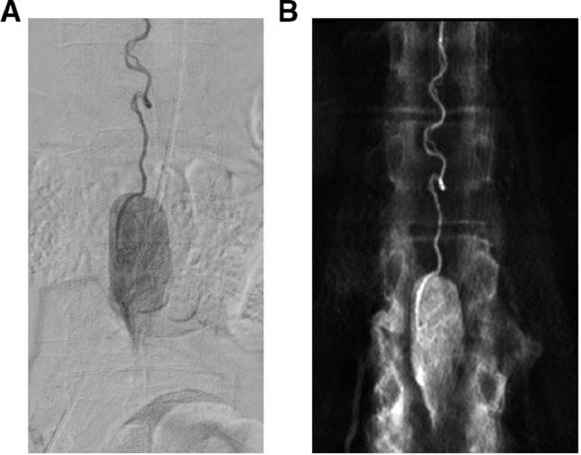
Selective spinal angiography via the left T12 intercostal artery. Conventional angiographic image (A), and CT reconstruction of 3D rotational angiography (B). Angiography revealed a highly vascular mass supplied by the anterior spinal artery (artery of Adamkiewicz). The lesión presented well defined margins and tumor stain in the late angiographic phase, like the appearance of a “silk cocoon.”

Intraoperative picture of the lesion, after durotomy (cranial to caudal from right to left). Hypervascular intradural extramedullary and well-marginated mass was observed.
References
-
- Miller C.A., Torack R.M. Secretory ependymoma of the filum terminale. Acta Neuropathol. 1970;15:240–250. - PubMed
-
- Gelabert-Gonzalez M. Paragangliomas of the lumbar region. Report of two cases and review of the literature. Neurosurg Spine. 2005;2(3):354–365. - PubMed
-
- Sonneland P.R.L., Scheithauer B.W., Lechago J., Crawford B.G., Onofrio B.M. Paraganglioma of the cauda equina region: clinico-pathologic study of 31 cases with special reference to immunocytology and ultrastructure. Cancer. 1986;15:1720–1735. - PubMed
-
- Yang C., Li G., Fang J., Wu L., Yang T., Deng X., Xu Y. Clinical characteristics and surgical outcomes of primary spinal paragangliomas. J Neurooncol. 2015:539–547. - PubMed
-
- Moran C.A., Rush W., Mena H. Primary spinal paragangliomas: a clinicopathological and immunohistochemical study of 30 cases. Histopathology. 1997;31(2):167–173. - PubMed
Publication types
LinkOut - more resources
Full Text Sources
