. 2019 Sep;13(3):031001-310017.
doi: 10.1115/1.4043461.

An Extensible Orthopaedic Wire Navigation Simulation Platform

Affiliations

An Extensible Orthopaedic Wire Navigation Simulation Platform

Steven Long et al. J Med Device. 2019 Sep.

Abstract

The demand for simulation-based skills training in orthopaedics is steadily growing. Wire navigation, or the ability to use 2D images to place an implant through a specified path in bone, is an area of training that has been difficult to simulate given its reliance on radiation based fluoroscopy. Our group previously presented on the development of a wire navigation simulator for a hip fracture module. In this paper, we present a new methodology for extending the simulator to other surgical applications of wire navigation. As an example, this paper focuses on the development of an iliosacral wire navigation simulator. We define three criteria that must be met to adapt the underlying technology to new areas of wire navigation; surgical working volume, system precision, and tactile feedback. The hypothesis being that techniques which fall within the surgical working volume of the simulator, demand a precision less than or equal to what the simulator can provide, and that require the tactile feedback offered through simulated bone can be adopted into the wire navigation module and accepted as a valid simulator for the surgeons using it. Using these design parameters, the simulator was successfully configured to simulate the task of drilling a wire for an iliosacral screw. Residents at the University of Iowa successfully used this new module with minimal technical errors during use.


Figures

Figure 1.
Figure 1.
A surgical wire drills into a plastic bone located just inside the opening in the tan, soft-tissue covering. The bone is mounted on the wire navigation simulator. A laptop displays artificial fluoroscope images used for training hip wire navigation. A camera system inside the simulator tracks the wire relative to the fixed bone to create artificial fluoroscopic images showing the wire position in bone.
Figure 2.
Figure 2.
The dimensions of the working volume are shown here. The ruler is positioned against the soft tissue which encloses the bone of the simulator. The space beyond the soft tissue defines the region of the image in which the wire can be seen by the camera system to calculate the wire position in bone.
Figure 3.
Figure 3.
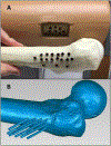
(A) The copper tubes used to create reproducible wire positions are shown here. The tubes are cemented in bone to create a reproducible slot that a wire can be inserted to for testing. (B) A laser scan of one of the bones with the wire positions for each slot. The laser scan defined the wire trajectory and position for each copper tube.
Figure 4.
Figure 4.
This shows the potential working volume for the iliosacral screw placement on the simulator. The area in dark blue defines the potential wire regions that would be used to successfully guide a wire into the iliosacral corridor.
Figure 5.
Figure 5.
These oblique views show the pelvic bone mounted to this simulator base. The soft tissue is not shown here. The black 3D printed materials act as a vice to hold the pelvic model in place while residents drill into the bone.
Figure 6.
Figure 6.
The three views used in the iliosacral procedure are shown here. The inlet and outlet views are taken at oblique angles to a patient, and the lateral view is taken directly along the corridor of the iliosacral joint.
Figure 7.
Figure 7.
Starting views in the bubble game shown here. In the game cartoon depictions of the pelvic anatomy were used to make the targets more clear to the trainees. The blue circles act as targets for the residents to pierce with their wire during the training exercise.
Figure 8.
Figure 8.
Errors of the wire tip position, after projecting into bone for different wire positions. Average tip errors at each position are indicated by color: green for errors < 1mm, yellow for errors between 1 and 2mm, blue for errors between 2 and 3mm, and red for errors > 3mm.
Figure 9.
Figure 9.
This graph illustrates the strong relationship between a resident’s performance on the target practice in the outlet view with the 3D target practice. Each residents’ performance on the outlet view bubble task is graphed on the y axis and the corresponding score on the 3D bubble task is shown on the x-axis. A strong correlation of R2 = 0.79 can be seen between these two metrics.

Similar articles

Cited by

References

    1. ACGME Highlights Its Standards on Resident Duty Hours. Available from: http://www.acgme.org/acgmeweb/tabid/363/Publications/Papers/PositionPape.... Accessed 5/20/2018
    1. ACGME Program Requirements for Graduate Medical Education in Orthopaedic Surgery. https://www.acgme.org/Portals/0/PFAssets/ProgramRequirements/260_orthopa.... Accessed 5/20/2018
    1. Ferguson PC, Kraemer W, Nousiainen M, Safir O, Sonnadara R, Alman B, Reznick R. Three-Year experience with an innovative, modular competency-based curriculum for orthopaedic training. J Bone Joint Surg Am. 2013. November 95 (21)e166. - PubMed
    1. Khanduja V, Lawrence JE, and Audenaert E, Testing the Construct Validity of a Virtual Reality Hip Arthroscopy Simulator. Arthroscopy, 2017. 33(3): p. 566–571. - PubMed
    1. Lopez G, et al., Construct Validity for a Cost-effective Arthroscopic Surgery Simulator for Resident Education. Journal of the American Academy of Orthopaedic Surgeons, 2016. 24(12): p. 886–894. - PubMed

LinkOut - more resources