. 2019 Jul 17:9:255.
doi: 10.3389/fcimb.2019.00255. eCollection 2019.

The Interaction of Klebsiella pneumoniae With Lipid Rafts-Associated Cholesterol Increases Macrophage-Mediated Phagocytosis Due to Down Regulation of the Capsule Polysaccharide

Affiliations

The Interaction of Klebsiella pneumoniae With Lipid Rafts-Associated Cholesterol Increases Macrophage-Mediated Phagocytosis Due to Down Regulation of the Capsule Polysaccharide

Miguel A Ares et al. Front Cell Infect Microbiol. .

Abstract

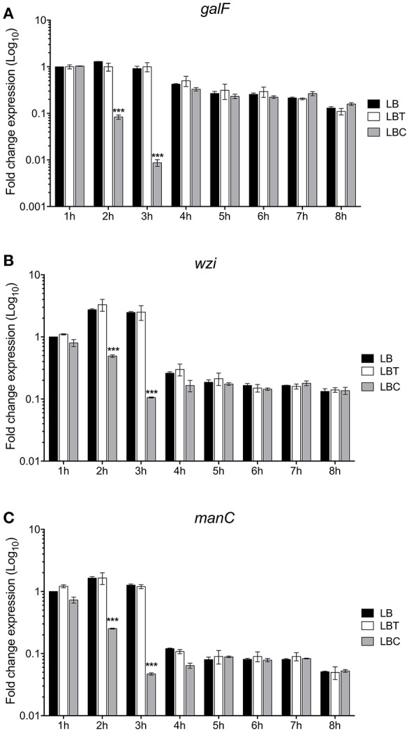
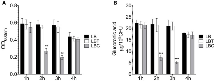
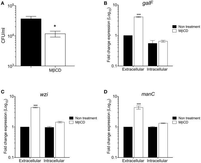
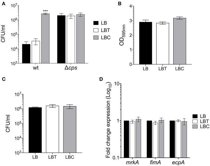
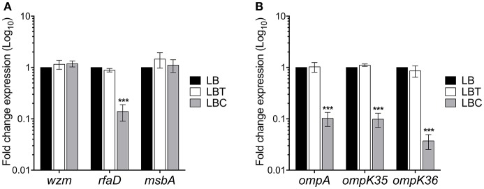
Klebsiella pneumoniae successfully colonizes host tissues by recognizing and interacting with cholesterol present on membrane-associated lipid rafts. In this study, we evaluated the role of cholesterol in the expression of capsule polysaccharide genes of K. pneumoniae and its implication in resistance to phagocytosis. Our data revealed that exogenous cholesterol added to K. pneumoniae increases macrophage-mediated phagocytosis. To explain this event, the expression of capsular galF, wzi, and manC genes was determined in the presence of cholesterol. Down-regulation of these capsular genes occurred leading to increased susceptibility to phagocytosis by macrophages. In contrast, depletion of cholesterol from macrophage membranes led to enhanced expression of galF, wzi, and manC genes and to capsule production resulting in resistance to macrophage-mediated phagocytosis. Cholesterol-mediated repression of capsular genes was dependent on the RcsA and H-NS global regulators. Finally, cholesterol also down-regulated the expression of genes responsible for LPS core oligosaccharides production and OMPs. Our results suggest that cholesterol plays an important role for the host by reducing the anti-phagocytic properties of the K. pneumoniae capsule facilitating bacterial engulfment by macrophages during the bacteria-eukaryotic cell interaction mediated by lipid rafts.

Keywords: H-NS; Klebsiella pneumoniae; RcsA; capsule; cholesterol; phagocytosis.


Figures

Figure 1
Figure 1
Cholesterol does not affect the K. pneumoniae growth. Growth curves of K. pneumoniae in LB medium with no supplement (LB), 0.05% tyloxapol (LBT), and 0.05% tyloxapol plus 50 μM cholesterol (LBC). Bacterial cultures were grown at 37°C for 8 h.
Figure 2
Figure 2
Cholesterol represses the transcription of capsular genes in K. pneumoniae. Fold change expression (RT-qPCR) of galF (A), wzi (B), and manC (C) genes. K. pneumoniae was grown in LB with no supplement (LB), 0.05% tyloxapol (LBT), and 0.05% tyloxapol plus 50 μM cholesterol (LBC). Bacterial cultures were grown at 37°C for 8 h. 16S rRNA was used as a reference gene for normalization. Data represent the mean of three independent experiments performed in triplicates. Statistically significant with respect to the WT bacteria grown in LB medium ***p < 0.001.
Figure 3
Figure 3
Cholesterol affects the production of capsular polysaccharide in K. pneumoniae. (A) Mucoviscosity of K. pneumoniae grown in LB with no supplement (LB), 0.05% tyloxapol (LBT), and 0.05% tyloxapol plus 50 μM cholesterol (LBC). The mucoviscosity was determined by low speed centrifugation and is expressed as OD600nm of the supernatant. (B) Capsule quantification by determination of the glucuronic acid concentration from capsular polysaccharides. Statistically significant with respect to the WT bacteria grown in ** p < 0.01 between LB medium and ***p < 0.001.
Figure 4
Figure 4
The depletion of cholesterol from macrophages lipids rafts increases the K. pneumoniae capsule polysaccharide. (A) Comparison of phagocytic uptake of K. pneumoniae by THP-1 macrophages treated without/with MβCD (methyl-β-cyclodextrin) to deplete cholesterol. Fold change expression (RT-qPCR) of galF (B), wzi (C), and manC (D) genes of K. pneumoniae (extra and intracellular) during the phagocytosis by THP-1 macrophages treated without/with MβCD. Data represent the mean of at least three independent experiments performed in triplicates (mean ± SD). Statistically significant with respect to the WT bacteria grown in LB medium * p < 0.05; *** p < 0.001.
Figure 5
Figure 5
Cholesterol enhances macrophage-mediated phagocytosis of K. pneumoniae. (A) Comparison of phagocytic uptake of K. pneumoniae grown under the indicated different conditions by THP-1 macrophages. A Δcps mutant was evaluated as control of phagocytosis. (B) Quantification of biofilm formation by measuring Crystal Violet uptake of K. pneumoniae in LB, LBT and LBC broths. (C) Adherence levels of K. pneumoniae grown in LB, LBT and LBC broths to A549 cells. (D) Fold change expression (RT-qPCR) of fimbrial genes. 16S rRNA was used as a reference gene for normalization. Data represent the mean of at least three independent experiments performed in triplicates (mean ± SD). Statistically significant with respect to the WT bacteria grown in LB medium ***p < 0.001.
Figure 6
Figure 6
RcsA and H-NS are involved in the cholesterol-mediated capsule repression. Fold change expression (RT-qPCR) of galF (A), wzi (B), and manC (C) genes in the presence of cholesterol [LB with no supplement (LB), 0.05% tyloxapol (LBT), and 0.05% tyloxapol plus 50 μM cholesterol (LBC)] in different backgrounds: wild-type, ΔrcsA and Δhns. 16S rRNA was used as a reference gene for normalization. (D) Effect of cholesterol on phagocytic uptake of K. pneumoniae wild-type, ΔrcsA, and Δhns by THP-1 macrophages. Data represent the mean of at least three independent experiments performed in triplicates (mean ± SD). Statistically significant with respect to the WT bacteria grown in LB medium ***p < 0.001.
Figure 7
Figure 7
Cholesterol affects other virulence genes in K. pneumoniae. Fold change expression (RT-qPCR) of genes responsible for LPS production (A) and outer membrane proteins (B). K. pneumoniae was grown in LB with no supplement (LB), 0.05% tyloxapol (LBT), and 0.05% tyloxapol plus 50 μM cholesterol (LBC) at 37°C for 3 h. 16S rRNA was used as a reference gene for normalization. Data represent the mean of three independent experiments performed in triplicates. Statistically significant with respect to the WT bacteria grown in LB medium ***p < 0.001.

References

    1. Alcantar-Curiel M. D., Blackburn D., Saldana Z., Gayosso-Vazquez C., Iovine N. M., De la Cruz M. A., et al. (2013). Multi-functional analysis of Klebsiella pneumoniae fimbrial types in adherence and biofilm formation. Virulence 4, 129–138. 10.4161/viru.22974 - DOI - PMC - PubMed
    1. Alcantar-Curiel M. D., Giron J. A. (2015). Klebsiella pneumoniae and the pyogenic liver abscess: implications and association of the presence of rpmA genes and expression of hypermucoviscosity. Virulence 6, 407–409. 10.1080/21505594.2015.1030101 - DOI - PMC - PubMed
    1. Allen-Vercoe E., Waddell B., Livingstone S., Deans J., Devinney R. (2006). Enteropathogenic Escherichia coli Tir translocation and pedestal formation requires membrane cholesterol in the absence of bundle-forming pili. Cell Microbiol. 8, 613–624. 10.1111/j.1462-5822.2005.00654.x - DOI - PubMed
    1. Ares M. A., Alcantar-Curiel M. D., Jimenez-Galicia C., Rios-Sarabia N., Pacheco S., De la Cruz M. A. (2013). Antibiotic resistance of gram-negative bacilli isolated from pediatric patients with nosocomial bloodstream infections in a Mexican tertiary care hospital. Chemotherapy 59, 361–368. 10.1159/000362085 - DOI - PubMed
    1. Ares M. A., Fernandez-Vazquez J. L., Rosales-Reyes R., Jarillo-Quijada M. D., Von Bargen K., Torres J., et al. (2016). H-NS nucleoid protein controls virulence features of Klebsiella pneumoniae by regulating the expression of type 3 pili and the capsule polysaccharide. Front. Cell Infect. Microbiol. 6:13. 10.3389/fcimb.2016.00013 - DOI - PMC - PubMed

MeSH terms