. 2019 Jul 16:6:40.
doi: 10.3389/fsurg.2019.00040. eCollection 2019.

Interoptic, Trans-lamina Terminalis, Opticocarotid Triangle, and Caroticosylvian Windows From Mini-Supraorbital, Frontomedial, and Pterional Perspectives: A Comparative Cadaver Study With Artificial Lesions

Affiliations

Interoptic, Trans-lamina Terminalis, Opticocarotid Triangle, and Caroticosylvian Windows From Mini-Supraorbital, Frontomedial, and Pterional Perspectives: A Comparative Cadaver Study With Artificial Lesions

Sasan Darius Adib et al. Front Surg. .

Abstract

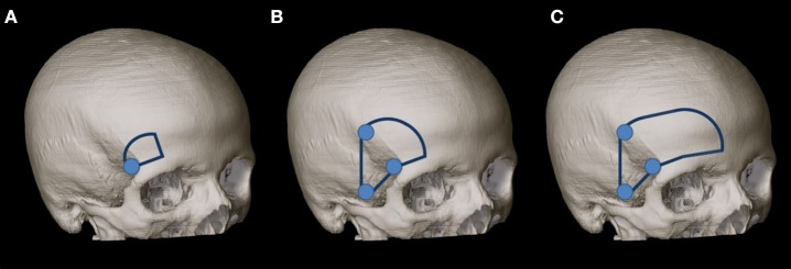
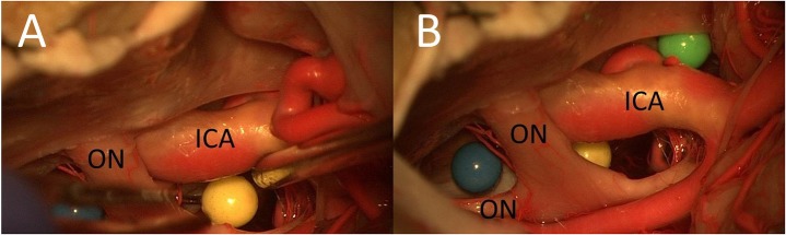
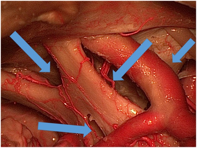
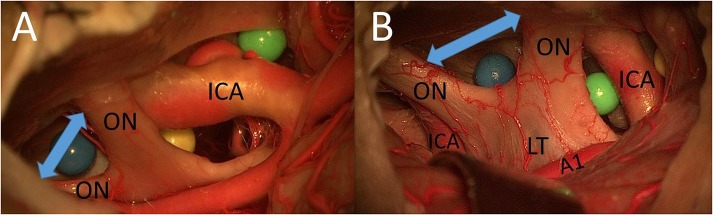
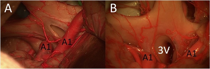
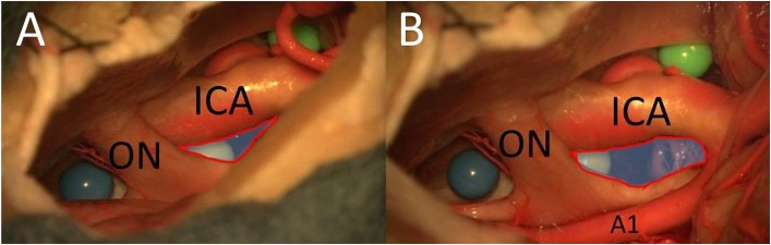
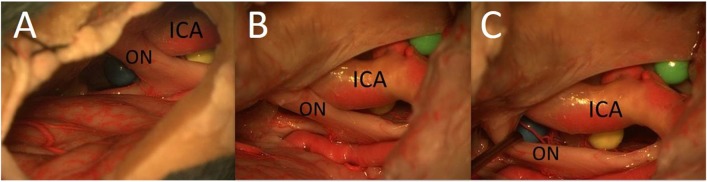
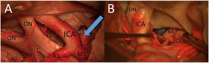
Introduction: The mini-supraorbital (MSO) and pterional (PT) approaches have been compared in a number of studies focusing on the treatment of aneurysms, craniopharyngiomas, and meningiomas. The goal of this study was to analyze the surgical exposure to different artificial lesions through interoptic (IO), trans-lamina terminalis (TLT), opticocarotid triangle (OCT), and caroticosylvian (CS) windows from the MSO, frontomedial (FM), and PT perspectives. Methods: The MSO, PT, and FM approaches were performed sequentially in two fixed cadaver heads. Three colored spheres were placed around the optic chiasm: (1) between the optic nerves; (2) between the optic nerve and the internal carotid artery; and (3) between the internal carotid artery and the oculomotor nerve. The surgical exposures to these structures by using the IO, TLT, OCT, and CS windows were compared. Results: (1) IO window: from the MSO and PT approaches, the total surgical exposure mainly allows visualization of contralateral lesions. The FM approach was superior for exploration of both sides of the area between the optic nerves. (2) TLT pathway: the MSO and PT approaches mainly expose the contralateral third ventricle wall. (3) OCT window: the PT approach allows exposure of a larger part of the sphere between the optic nerve and the internal carotid artery than the MSO approach. (4) CS window: the PT approach allows a better exposure of lateral structures such as the oculomotor nerve and of the medial prepontine area in comparison to the MSO approach. Conclusion: Simulation of the surgical situation with artificial lesions is a good model for comparing surgical perspectives and for analyzing feasibility of lesion exposure and resection.

Keywords: artificial lesions; caroticosylvian window; interoptic window; opticocarotid triangle window; trans-lamina terminalis approach.


Figures

Figure 1
Figure 1
(A) MSO craniotomy was performed according to Reisch et al. (2) with a 2 × 2.5-cm extension. (B) Second, the MSO approach was extended to a PT approach according to Yaşargil et al. (21) with a size of 4 × 6 cm. (C) The craniotomy was enlarged (as our third step) for the exposure to the frontal midline.
Figure 2
Figure 2
Placement of artificial lesions (three colored spheres) in key locations (A,B) (ON, optic nerve).
Figure 3
Figure 3
Various ways (blue arrows) of exposing the area under and around the optic chiasm and through the lamina terminalis to the third ventricle.
Figure 4
Figure 4
(A) Cadaver 1: IO window from PT perspective. (B) Cadaver 2: IO window from unilateral FM approach (ON, optic nerve; A1, proximal segment of anterior cerebral artery; LT, lamina terminalis).
Figure 5
Figure 5
(A) MSO and PT approaches allowed access to contralateral lesions of the third ventricle through the lamina terminalis. (B) The FM approach was the best way to reach deep lesions of the third ventricle through the lamina terminalis (3V, third ventricle; A1, proximal segment of anterior cerebral artery; LT, lamina terminalis).
Figure 6
Figure 6
The opticocarotid window from MSO (A) and PT approaches (B) (blue area with red frame = area of exposure of the OCT) (ON, optic nerve; A1, proximal segment of anterior cerebral artery).
Figure 7
Figure 7
The area under the optic chiasm: it is not possible to reach the area under the optic chiasm from the lateral aspect using the MSO approach (A); the area under the optic chiasm with the undersurface of the chiasm is easily visualized from lateral perspective through the opticocarotid window using the PT approach (B,C) (ON, optic nerve).
Figure 8
Figure 8
(A) CS window from PT perspective. (B) The prepontine area through the caroticosylvian window using the PT approach: mobilization of the ICA was necessary to increase the view to the BA, the PCA, the rami ad pontem, and the anterior upper surface of the brainstem (ON, optic nerve; A1, proximal segment of anterior cerebral artery; M1, proximal segment of middle cerebral artery).

References

    1. Figueiredo EG, Deshmukh V, Nakaji P, Deshmukh P, Crusius N, Spetzler RF, et al. . An anatomical evaluation of the mini-supraorbital approach and comparison with standard craniotomies. Neurosurgery. (2006) 59:212–20. 10.1227/01.NEU.0000223365.55701.F2 - DOI - PubMed
    1. Reisch R, Perneczky A, Filippi R. Surgical technique of the supraorbital key-hole craniotomy. Surg Neurol. (2003) 59:223–7. 10.1016/S0090-3019(02)01037-6 - DOI - PubMed
    1. van Lindert E, Perneczky A, Fries G, Pierangeli E. The supraorbital keyhole approach to supratentorial aneurysms: concept and technique. Surg Neurol. (1998) 49:481–90. 10.1016/S0090-3019(96)00539-3 - DOI - PubMed
    1. Yasargil MG, Fox JL. The microsurgical approach to intracranial aneurysms. Surg Neurol. (1975) 3:7–14. - PubMed
    1. Chalouhi N, Jabbour P, Ibrahim I, Starke RM, Younes P, El Hage G, et al. . Surgical treatment of ruptured anterior circulation aneurysms: comparison of pterional and supraorbital keyhole approaches. Neurosurgery. (2013) 72:437–42. 10.1227/NEU.0b013e3182804e9c - DOI - PubMed

LinkOut - more resources