Review
. 2019 Jun;5(Suppl 1):S41-S56.
doi: 10.21037/jss.2019.04.19.

The benefit zone of full-endoscopic spine surgery

Affiliations
Review

The benefit zone of full-endoscopic spine surgery

Saqib Hasan et al. J Spine Surg. 2019 Jun.

Abstract

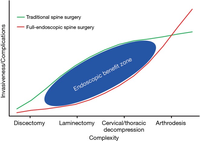
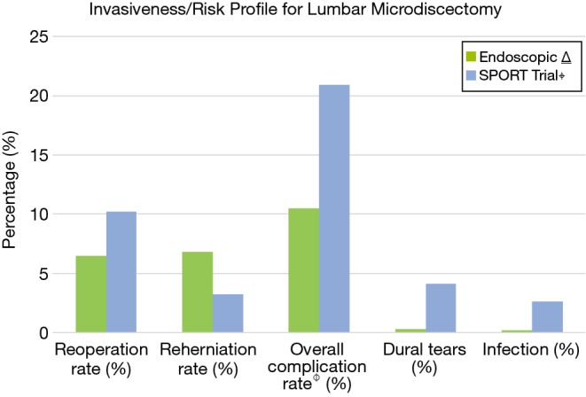
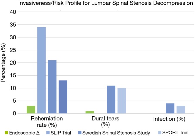
Minimally invasive spine procedures have undergone rapid development during the last decade. Efforts to decrease muscle crush injuries during prolonged retraction, avoid significant soft tissue stripping and minimize bony resection are surgical principles that are employed to prevent iatrogenic instability and provide patients with decreased post-operative pain and disability. Full-endoscopic spine surgery represents a tool for the spine surgeon to provide targeted access to spinal pathology utilizing these principles. Endoscopic techniques have seen over 30 years of evolution and innovation, however, early iterations of these techniques largely focused on transforaminal lumbar microdiscectomies. Currently, endoscopic techniques are utilized for approaching pathology in the cervical, thoracic and lumbar spine. There has been a growing body of literature that not only confirms the efficacy of these procedures but also underscores the advantages these procedures offer with respect to less morbidity and safer complication profiles. Endoscopic decompressions have been utilized in the settings of degenerative spinal stenosis, spondylolisthesis, scoliosis, previous fusion, tumor and infection. Furthermore, endoscopic interbody fusion has also been utilized in the lumbar spine as technology continues to advance. As technological innovation continues to facilitate reproducible surgical technique and expand the indications for use, we believe that endoscopic spine surgical techniques will provide surgeons with a more powerful and less morbid approach to spinal pathology that ultimately elevates the standard of care when treating our patients. We present a brief review of the history of endoscopic spine surgery, an overview of current techniques and review current outcomes of endoscopic spine surgical procedures in the context of an invasiveness/complexity index to elucidate the benefit zone of these newer techniques.

Keywords: Endoscopic spine surgery; invasiveness index; minimally invasive spine surgery.


Conflict of interest statement

Conflicts of Interest: CP Hofstetter: consulting and teaching for J&J, Globus and Joimax; R Hârtl: consultant brainlab DPS. S Hasan has no conflicts of interest to declare.

Figures

Figure 1
Figure 1
(A) Depicts a sagittal cross-sectional image of a cadaveric specimen illustrating Kambin’s triangle (K); (B) depicts a sagittal cross-sectional image from a T2-weighted MRI illustrating Kambin’s triangle (K). The hypotenuse of the triangle is the exiting nerve root, the base (width) is the superior border of the caudal vertebra, and the height is the dura/traversing nerve root. SAP represents the superior articular process of the caudal vertebrae, ID represents the intervertebral disc, ENR depicts the exiting nerve root and the gray arrows depict the contents of the foramen including perineural fat, perineural vessels and foraminal ligaments.
Figure 2
Figure 2
(A) Depicts an axial cross-sectional image from a T2-weighted MRI of the sub-axial cervical spine. The red arrows indicate potential approach corridors for the interlaminar approach which allow dorsal decompression of ipsilateral foraminal stenosis or central stenosis; (B) depicts an axial cross-sectional image from a T2-weighted MRI of the lumbar spine. The red arrows indicate potential approach corridors for the interlaminar approach which allow decompression of central stenosis and/or ipsilateral and contralateral lateral recess stenosis. The yellow arrows indicate potential approach corridors for the transforaminal approach which allow decompression of ipsilateral extra-foraminal, foraminal and lateral recess stenosis. Note that for the thoracic spine, the interlaminar approach corridor is more limited than the lumbar spine due to the presence of the spinal cord.
Figure 3
Figure 3
(A) This is an axial cross-sectional CT image of the L2/L3 level status-post left-sided endoscopic unilateral laminotomy for bilateral decompression (ULBD) for lumbar spinal stenosis. The endoscope allows for excellent ipsilateral facet joint preservation given off-angle visualization and the ability to tilt the small diameter endoscope out into the lateral recess; (B) an endoscopic picture depicts the dural sac after full-endoscopic decompression of the L2/3 level via a unilateral laminotomy for bilateral decompression (ULBD). The white star represents the central portion of the decompressed dural sac. The red stars represent the decompressed traversing nerve roots. The R on the compass represents rostral, and the C represents caudal.
Figure 4
Figure 4
The surgical invasiveness and complexity index is a qualitative graphical representation which plots the perceived complexity and invasiveness of spine surgical procedures which can be accomplished via open, minimally-invasive tubular or endoscopic methods. The endoscopic benefit zone is depicted by the blue ellipse and represents the theorized advantage endoscopic techniques have for different surgical procedures based on comparative surgical risk profiles from current published data. The advantage of endoscopic techniques is more pronounced for moderate to higher complexity procedures such as decompression for lumbar spinal stenosis and cervical/thoracic decompression.
Figure 5
Figure 5
The graph depicts the invasiveness and risk profiles for endoscopic versus open microdiscectomy based on current published data. Endoscopic ⍙: data from this cohort was obtained from aggregate data from 5 prospective trials listed in Table 1. SPORT Trial⌖: data from this cohort was obtained from Weinstein et al. (43). Overall complication rate : the overall complication rate includes all reported peri-operative events and complications including reoperation.
Figure 6
Figure 6
The graph depicts the invasiveness and risk profiles for endoscopic decompression versus open laminectomy for lumbar spinal stenosis based on current published and available data. Data from the SLIP trial was obtained from Ghogawala et al. (64); the number of patients with dural tears and infection were not reported. Data from the Swedish Spinal Stenosis Study was obtained from Forsth et al. (65). Data from the SPORT trial was obtained from Weinstein et al. (57,62). Endoscopic ⍙: data from the endoscopic cohort was obtained from our institutional data (unpublished).
Figure 7
Figure 7
An endoscopic picture depicts C5 nerve root after full-endoscopic right-sided foraminotomy of the C4/5 level. The white star represents the spinal cord. The decompressed C5 nerve root (red star) has been decompressed passed the rostral pedicle (P). A disc fragment was removed from the disc space (D) in the axilla of the nerve root. The R on the compass represents rostral, and the C represents caudal.
Figure 8
Figure 8
(A) An endoscopic picture depicts a lumbar synovial cyst (red star) abutting the thecal sac and traversing nerve root (white star). A high-speed endoscopic burr was used to create a laminotomy at the inferior articular process (IAP) and a portion of the ligamentum flavum (FL) was removed; (B) an endoscopic picture depicts revision decompression following placement of an interlaminar spacer. An endoscopic high-speed burr and Kerrison ronguers were used to create expand the previous laminotomy and remove a portion of the implant (H) and dissect through scar tissue (S). Following resection of the ligamentum flavum (FL), the thecal sac is identified (white star) and subsequently decompressed. The R on the compass represents rostral, and the C represents caudal.

References

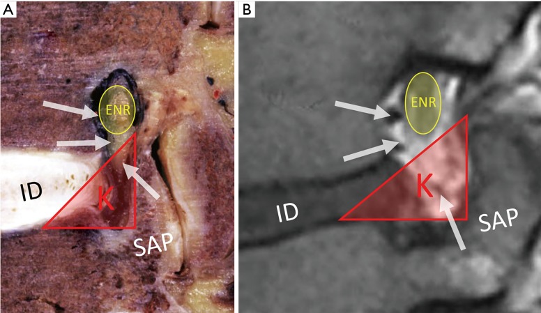
    1. Kambin P, Brager MD. Percutaneous posterolateral discectomy. Anatomy and mechanism. Clin Orthop Relat Res 1987;(223):145-54. - PubMed
    1. Gibson JNA, Subramanian AS, Scott CEH. A randomised controlled trial of transforaminal endoscopic discectomy vs microdiscectomy. Eur Spine J 2017;26:847-56. 10.1007/s00586-016-4885-6 - DOI - PubMed
    1. Hoogland T, Schubert M, Miklitz B, et al. Transforaminal posterolateral endoscopic discectomy with or without the combination of a low-dose chymopapain: a prospective randomized study in 280 consecutive cases. Spine (Phila Pa 1976) 2006;31:E890-7. 10.1097/01.brs.0000245955.22358.3a - DOI - PubMed
    1. Komp M, Hahn P, Oezdemir S, et al. Bilateral spinal decompression of lumbar central stenosis with the full-endoscopic interlaminar versus microsurgical laminotomy technique: a prospective, randomized, controlled study. Pain Physician 2015;18:61-70. - PubMed
    1. Ruetten S, Komp M, Merk H, et al. Surgical treatment for lumbar lateral recess stenosis with the full-endoscopic interlaminar approach versus conventional microsurgical technique: a prospective, randomized, controlled study. J Neurosurg Spine 2009;10:476-85. 10.3171/2008.7.17634 - DOI - PubMed