Review
. 2019 Jun;5(Suppl 1):S74-S83.
doi: 10.21037/jss.2019.04.21.

MIS approaches in the cervical spine

Affiliations
Review

MIS approaches in the cervical spine

Ibrahim Hussain et al. J Spine Surg. 2019 Jun.

Abstract

Minimally invasive surgical approaches for the treatment of spinal pathologies have accelerated over the past three decades and resulted in superior functional outcomes with less complications. Yet cervical pathologies have been slower to gain traction for multiple anatomical factors and its "high-risk" profile. Various minimally invasive techniques for cervical disease have now been described and validated in long-term studies with comparable outcomes to traditional open approaches and concomitant reduction in morbidity and socioeconomic costs. Transnasal operations can be used to treat ventral upper cervical disease, circumventing traditional and morbid transoral approaches. Posterior-based focused treatments for radiculopathy and myelopathy such as tubular-guided foraminotomies and unilateral laminotomies for bilateral cord decompression have also been described and becoming increasingly less invasive. Cervical fusions can now be performed percutaneously through modified, stand-alone facet joint cages that can be packed with allogeneic bone graft. These advances have been facilitated by the development of intraoperative imaging technologies (intraoperative CT) and 3-dimensional stereotactic navigation software. While this review focuses on these procedures and evidence-based outcomes data, the future for MIS applications in cervical spine surgery will continue to evolve over the coming years with wider indications and technological adjuncts.

Keywords: 3D; cervical; endonasal; endoscopic; facet cage; foraminotomy; fusion; interfacet; laminectomy; laminotomy; minimally invasive spine (MIS); navigation; odontoidectomy; over the top; posterior.


Conflict of interest statement

Conflicts of Interest: The authors have no conflicts of interest to declare.

Figures

Figure 1
Figure 1
Pre- and postoperative imaging status post endonasal odontoidectomy. (A) Preoperative T1-weighted MR image of a retroflexed odontoid with basilar invagination and kinking of the cervicomedullary junction in a patient with Type 1 Chiari malformation; (B) postoperative T1-weighted MR image following odontoidectomy with marked improvement in cervicomedullary kniking; (C) preoperative and (D) postoperative CT images demonstrating the bony anatomy before and after resection of the anterior arch of C1 and odontoidectomy. Note the occipitocervical fusion hardware which preceded the endonasal odontoidectomy.
Figure 2
Figure 2
Endoscopic endonasal odontoidectomy. (A) Sagittal, and (B) axial intraoperative 3D navigation screenshots demonstrating the endonasal approach and trajectory to the odontoid; (C) intraoperative endoscopic photograph of the odontoid being drilled with an extended, fluted 4 mm diamond burr.
Figure 3
Figure 3
3D navigation intraoperative screenshots demonstrating the ipsilateral and contralateral foramen that can be safely accessed through a unilateral approach for performing posterior cervical foraminotomy.
Figure 4
Figure 4
Preoperative (A) sagittal and (B) axial T2 weighted MR images demonstrating severe C4/5 stenosis intramedullary hyperintensity indicative of myelomalacia. Note on axial cuts the complete loss of CSF signal around the spinal cord. Postoperative (C) sagittal and (D) axial cuts following minimally invasive tubular decompression via unilateral laminotomy for bilateral decompression (ULBD “over the top” technique). Note the bilateral laminar bony resection with restoration of CSF signal.
Figure 5
Figure 5
Intraoperative axial 3D navigation screenshots demonstrating the lateral extents of the unilateral laminotomy for bilateral decompression (ULBD “over the top” technique).
Figure 6
Figure 6
Sagittal (A) and (B,C,D) axial T2 MR image of patient with advanced cervical spondylosis and degenerative changes, with autofusion of C5/6 and severe canal stenosis at C4/5. (E) CT scan demonstrates severe degenerative facet joint changes including fluid and vacuum phenomenon (yellow arrow) suggesting instability. This patient has radiographic evidence of severe central stenosis and instability, therefore decompression and stabilization are indicated. However, the patient had multiple comorbidities, therefore a minimally invasive interfacet joint cage implant was used for fusion as opposed to lateral mass screw and rod construct in conjunction with ULBD. ULBD, unilateral laminotomy for bilateral decompression.
Figure 7
Figure 7
Intraoperative CT demonstrates the bony resection (yellow arrow) following ULBD with interfacet joint cage placements, which are in adequate position. ULBD, unilateral laminotomy for bilateral decompression.
Figure 8
Figure 8
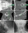
Intraoperative 3D navigation screenshots demonstrating the trajectory and ultimate target of the interfacet joint cage (A,B,C,D). Postoperative (E) lateral and (F) anteroposterior radiographs following bilateral interfacet joint cage implantation.

References

    1. Jain N, Verma R, Garga UC, et al. CT and MR imaging of odontoid abnormalities: A pictorial review. Indian J Radiol Imaging 2016;26:108-19. 10.4103/0971-3026.178358 - DOI - PMC - PubMed
    1. Klekamp J. Chiari I malformation with and without basilar invagination: a comparative study. Neurosurg Focus 2015;38:E12. 10.3171/2015.1.FOCUS14783 - DOI - PubMed
    1. Kahilogullari G, Eroglu U, Yakar F, et al. Endoscopic Endonasal Approaches to Craniovertebral Junction Pathologies: A Single-Center Experience. Turk Neurosurg 2018. [Epub ahead of print]. 10.5137/1019-5149.JTN.23781-18.2 - DOI - PubMed
    1. Shriver MF, Kshettry VR, Sindwani R, et al. Transoral and transnasal odontoidectomy complications: A systematic review and meta-analysis. Clin Neurol Neurosurg 2016;148:121-9. 10.1016/j.clineuro.2016.07.019 - DOI - PubMed
    1. Choi D, Crockard HA. Evolution of transoral surgery: three decades of change in patients, pathologies, and indications. Neurosurgery 2013;73:296-303; discussion 303-4. 10.1227/01.neu.0000430324.24623.10 - DOI - PubMed