

Patient-Specific Modeling of Heart Valves: From Image to Simulation
- PMID: 31380522
- PMCID: PMC6679972
- DOI: 10.1007/978-3-642-38899-6_17
Patient-Specific Modeling of Heart Valves: From Image to Simulation
Abstract
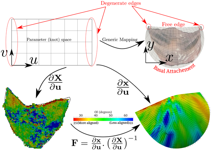
Heart valves play a very important role in the functioning of the heart and many of the heart failures are related to the valvular dysfunctions, e.g. aortic stenosis and mitral regurgitation. As the medical field is moving towards a patient-specific diagnosis and treatment procedures, modeling of heart valves with patient-specific information is becoming a significant tool in medical field. Here we present the ingredients for valve simulation specifically the aortic valve, with a main focus on a novel spline-based mapping technique which solves many issues in generating patient-specific models - the microstructural mapping, the pre-strain calculations, prescribing dynamic boundary conditions, validation and inverse-modeling to obtain material parameters.
Figures


References
-
- Rajamannan N: Cardiac Valvular Medicine. Springer; (2012)
-
- Cottrell J, Hughes T, Bazilevs Y: Isogeometric analysis: toward integration of CAD and FEA. Wiley; (2009)
-
- Ionasec RI, Voigt I, Georgescu B, Wang Y, Houle H, Vega-Higuera F, Navab N, Comaniciu D: Patient-specific modeling and quantification of the aortic and mitral valves from 4-d cardiac ct and tee. IEEE Transactions on Medical Imaging 29(9), 1636–1651 (2010) - PubMed
Grants and funding
LinkOut - more resources
Full Text Sources
Research Materials