. 2021 Mar 19;16(16):1342-1348.
doi: 10.4244/EIJ-D-19-00555.

Ultrasound-guided access to the distal radial artery at the anatomical snuffbox for catheter-based vascular interventions: a technical guide

Affiliations

Ultrasound-guided access to the distal radial artery at the anatomical snuffbox for catheter-based vascular interventions: a technical guide

Anastasia Hadjivassiliou et al. EuroIntervention. .

Abstract

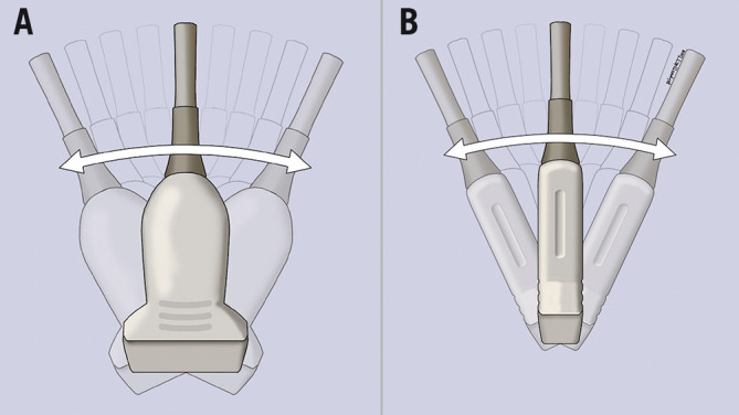
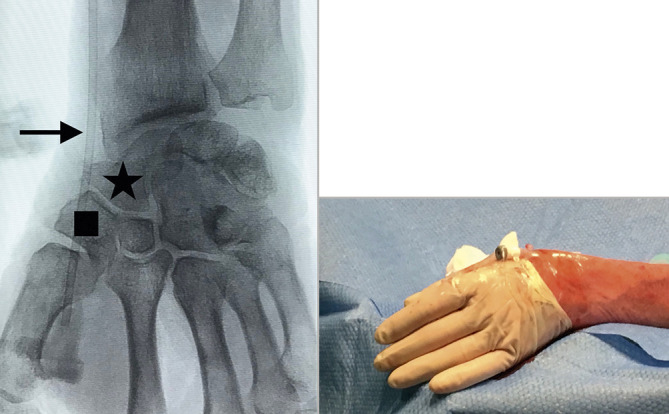
Conventional radial access has been shown to have many advantages over the transfemoral approach. The risk of potential radial artery occlusion and subsequent hand ischaemia can be reduced further by accessing the vessel distally at the anatomical snuffbox, allowing maintenance of antegrade flow to the hand by the superficial palmar arch branch. Additional potential advantages of distal radial access in comparison to the conventional radial approach at the wrist include fewer puncture-site complications and faster post-procedural haemostasis as the vessel is very superficial. Furthermore, it provides another safe, non-femoral option for vascular access. The use of ultrasound guidance enables the operator to identify important anatomical landmarks and avoid injuring adjacent structures. We provide a detailed step-by-step guide for performing distal radial access using sonographic and anatomical correlation, thereby facilitating safe access and optimising technical success.


Conflict of interest statement

S. Nathan is a consultant for Medtronic, Merit Medical and Terumo Interventional Systems. F. Kiemeneij is a consultant for Merit Medical. D. Klass is a consultant for Merit Medical. The other author has no conflicts of interest to declare.

Figures

Figure 1
Figure 1
Identification of basic US anatomy at the ventral aspect of the wrist for conventional radial arterial access. The radial artery (arrow). The distal radius (polygon) is represented by a bright line with posterior shadowing. The various soft tissues on the volar aspect of the wrist are seen as different shades on the greyscale image (asterisk).
Figure 2
Figure 2
US transducer probes. A curvilinear probe (A) emits a convex beam; the “hockey stick” probe (B) provides a comparatively small field of view. The linear transducer (C) emits a parallel beam and is ideal for vascular imaging.
Figure 3
Figure 3
Operator US scanning technique. A) Correct scanning technique with the operator’s hand partly resting on the patient and providing light pressure on soft tissues. B) & C) Incorrect handling of the US probe, resulting in an unstable probe position, excess compression and thus collapse of the radial artery.
Figure 4
Figure 4
Optimisation of US image. Image demonstrating the near (single asterisk) and far (double asterisk) fields during scanning. The depth has been adjusted so that the structures in the near field comprise the majority of the image. The key anatomical structures that should be identified in the anatomical snuffbox include the distal radial artery (R), cephalic vein branches (C), extensor pollicis longus (EPL), extensor pollicis brevis (EPB), abductor pollicis longus (APL) and scaphoid (S).
Figure 5
Figure 5
Arterial Doppler vascular assessment. US images of the distal radial artery (arrow) before and after Doppler tool is applied confirming patency.
Figure 6
Figure 6
Sonographic and anatomic correlation. A) – D) Different transducer positions and their corresponding US images starting distally at the first dorsal web space (A), moving proximally to the anatomical snuffbox over the scaphoid (D). Arrow: distal radial artery; circle: first dorsal interosseous muscle; filled triangle: thumb metacarpal; unfilled triangle: index metacarpal; square: trapezium bone; star: scaphoid bone.
Figure 7
Figure 7
Local anaesthetic skin infiltration. The probe should be held with the operator’s non-dominant hand and skin infiltration performed using the dominant hand.
Figure 8
Figure 8
Intravascular access. The position of the needle and wire at the anatomical snuffbox. Confirmation of appropriate intravascular position of the wire can be obtained on the US image as indicated by the arrows.
Figure 9
Figure 9
Sonographic vascular assessment. A) Heel-toe movement; the US probe is tilted on its long axis. B) Toggle movement; the US probe is tilted on its short axis.
Figure 10
Figure 10
Distal radial access sheath position. Fluoroscopic image showing the position of the vascular sheath. The puncture site is over the trapezium (square), with the sheath crossing over the scaphoid (star) and radioscaphoid joint. The location which is at the highest risk of kinking is indicated by the arrow. Once the sheath is inserted, it is secured as shown in the image.

References

    1. Kiemeneij F, Laarman GJ. Percutaneous transradial artery approach for coronary stent implantation. Cathet Cardiovasc Diagn. 1993;30:173–8. doi: 10.1002/ccd.1810300220. - DOI - PubMed
    1. Chase AJ, Fretz EB, Warburton WP, Klinke WP, Carere RG, Pi D, Berry B, Hilton JD. Association of the arterial access site at angioplasty with transfusion and mortality: the M.O.R.T.A.L study (Mortality benefit Of Reduced Transfusion after percutaneous coronary intervention via the Arm or Leg). Heart. 2008;94:1019–25. doi: 10.1136/hrt.2007.136390. - DOI - PubMed
    1. Jolly SS, Yusuf S, Cairns J, Niemelä K, Xavier D, Widimský P, Budaj A, Niemelä M, Valentin V, Lewis BS, Avezum A, Steg PG, Rao SV, Gao P, Afzal R, Joyner CD, Chrolavicius S, Mehta SR RIVAL trial group. Radial versus femoral access for coronary angiography and intervention in patients with acute coronary syndromes (RIVAL): a randomised, parallel group, multicentre trial. Lancet. 2011;377:1409–20. - PubMed
    1. Valgimigli M, Gagnor A, Calabro P, Frigoli E, Leonardi S, Zaro T, Rubartelli P, Briguori C, Ando G, Repetto A, Limbruno U, Cortese B, Sganzerla P, Lupi A, Galli M, Colangelo S, Ierna S, Ausiello A, Presbitero P, Sardella G, Varbella F, Esposito G, Santarelli A, Tresoldi S, Nazzaro M, Zingarelli A, de Cesare N, Rigattieri S, Tosi P, Palmieri C, Brugaletta S, Rao SV, Heg D, Rothenbuhler M, Vranckx P, Jüni P MATRIX Investigators. Radial versus femoral access in patients with acute coronary syndromes undergoing invasive management: a randomised multicentre trial. Lancet. 2015;385:2465–76. - PubMed
    1. Kiemeneij F. Left distal transradial access in the anatomical snuffbox for coronary angiography (ldTRA) and interventions (ldTRI). EuroIntervention. 2017;13:851–7. doi: 10.4244/EIJ-D-17-00079. - DOI - PubMed