Review
. 2019 Aug 5;129(10):4009-4021.
doi: 10.1172/JCI129190.

What lipodystrophies teach us about the metabolic syndrome

Review

What lipodystrophies teach us about the metabolic syndrome

Jake P Mann et al. J Clin Invest. .

Abstract

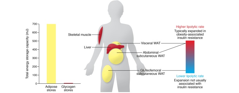
Lipodystrophies are the result of a range of inherited and acquired causes, but all are characterized by perturbations in white adipose tissue function and, in many instances, its mass or distribution. Though patients are often nonobese, they typically manifest a severe form of the metabolic syndrome, highlighting the importance of white fat in the "safe" storage of surplus energy. Understanding the molecular pathophysiology of congenital lipodystrophies has yielded useful insights into the biology of adipocytes and informed therapeutic strategies. More recently, genome-wide association studies focused on insulin resistance have linked common variants to genes implicated in adipose biology and suggested that subtle forms of lipodystrophy contribute to cardiometabolic disease risk at a population level. These observations underpin the use of aligned treatment strategies in insulin-resistant obese and lipodystrophic patients, the major goal being to alleviate the energetic burden on adipose tissue.


Conflict of interest statement

Conflict of interest: The authors have declared that no conflict of interest exists.

Figures

Figure 1
Figure 1. White adipose tissue contains the body’s major store of energy.
Even lean adults store 600–800 mJ of energy as triglyceride in adipose tissue compared with 6 to 8 mJ as glycogen in liver and muscle. The physiological regulation of triglyceride stores varies in different adipose tissue depots. Gluteofemoral subcutaneous white adipose is relatively insulin sensitive, and its expansion is not associated with cardiometabolic disease, whereas visceral and abdominal (upper body) subcutaneous adipose tissue has a higher rate of lipolysis and is more closely linked with insulin resistance. WAT, white adipose tissue.
Figure 2
Figure 2. Some of the genes/proteins in which mutations cause lipodystrophy have well-characterized roles in the function of adipocytes.
PPARγ (mutated in FPLD3) is the “master regulator” of adipogenesis. It heterodimerizes with retinoid X receptor and coordinates the transcription of multiple proteins central to adipocyte function (e.g., perilipin, CD36, and lipoprotein lipase). BSCL2, or seipin (mutated in CGL2), is an ER protein required for early lipid droplet (LD) biogenesis. AGPAT2 (mutated in CGL1) is necessary for the conversion of glycerophosphates (G-3-P) into triacylglycerols (TAGs) using fatty acids linked to coenzyme A (FA-CoA). CAV1 (mutated in CGL3) and PTRF (mutated in CGL4) are required for the formation of caveolae, which may be sites for nonesterified fatty acid (NEFA) uptake. PLIN1 (mutated in FPLD4) regulates lipolysis from lipid droplets, and HSL (mutated in FPLD6) is one of the lipases involved in this process. Finally, CIDEC (mutated in FPLD5) is required for the formation of unilocular lipid droplets, though how this is achieved is unclear.
Figure 3
Figure 3. The severity of lipodystrophy and the degree of adipose dysfunction correlate broadly with the severity of insulin resistance.
This principle extends from the most extreme form of lipodystrophy, congenital generalized lipodystrophy (CGL), through familial partial lipodystrophies (FPLDs) to the general population. Individuals in the highest quintile (Q5) for a polygenic risk score for insulin resistance (see Lotta et al.; ref. 175) have less gluteofemoral fat, resulting in an “apple-shaped” fat distribution, whereas those in the lowest quintile (Q1) manifest a protective “pear-shaped” fat distribution and are less insulin resistant. FPLD type 1 (FPLD1) represents an intermediate state between other monogenic forms of FPLD and the highest-risk individuals from the general population. The degree of genetic disruption of adipose tissue also correlates with these phenotypes, as exemplified by the impact of a range of PPARG mutations: complete loss of PPARγ function can cause CGL; dominant-negative PPARG mutations cause FPLD3; and common PPARG variants affect insulin resistance at a population level. Exemplars of PPARG mutations in each of these categories have been included. Each black dot represents a distinct monogenic disease (see Table 1 for classifications), and red diamonds schematically represent common genetic variants that influence adipogenesis and insulin resistance.

References

    1. Melvin A, O’Rahilly S, Savage DB. Genetic syndromes of severe insulin resistance. Curr Opin Genet Dev. 2018;50:60–67. doi: 10.1016/j.gde.2018.02.002. - DOI - PubMed
    1. Virtue S, Vidal-Puig A. Adipose tissue expandability, lipotoxicity and the Metabolic Syndrome — an allostatic perspective. Biochim Biophys Acta. 2010;1801(3):338–349. - PubMed
    1. Cuthbertson DJ, et al. What have human experimental overfeeding studies taught us about adipose tissue expansion and susceptibility to obesity and metabolic complications? Int J Obes (Lond) 2017;41(6):853–865. doi: 10.1038/ijo.2017.4. - DOI - PubMed
    1. Melvin A, et al. Roux-en-Y gastric bypass surgery in the management of familial partial lipodystrophy type 1. J Clin Endocrinol Metab. 2017;102(10):3616–3620. doi: 10.1210/jc.2017-01235. - DOI - PMC - PubMed
    1. Grundfest-Broniatowski S, Yan J, Kroh M, Kilim H, Stephenson A. Successful treatment of an unusual case of FPLD2: the role of Roux-en-Y gastric bypass-case report and literature review. J Gastrointest Surg. 2017;21(4):739–743. doi: 10.1007/s11605-016-3300-2. - DOI - PubMed

Publication types