. 2019 Aug 5;15(8):e1007962.
doi: 10.1371/journal.ppat.1007962. eCollection 2019 Aug.

Convergent evolution in the mechanisms of ACBD3 recruitment to picornavirus replication sites

Affiliations

Convergent evolution in the mechanisms of ACBD3 recruitment to picornavirus replication sites

Vladimira Horova et al. PLoS Pathog. .

Abstract

Enteroviruses, members of the family of picornaviruses, are the most common viral infectious agents in humans causing a broad spectrum of diseases ranging from mild respiratory illnesses to life-threatening infections. To efficiently replicate within the host cell, enteroviruses hijack several host factors, such as ACBD3. ACBD3 facilitates replication of various enterovirus species, however, structural determinants of ACBD3 recruitment to the viral replication sites are poorly understood. Here, we present a structural characterization of the interaction between ACBD3 and the non-structural 3A proteins of four representative enteroviruses (poliovirus, enterovirus A71, enterovirus D68, and rhinovirus B14). In addition, we describe the details of the 3A-3A interaction causing the assembly of the ACBD3-3A heterotetramers and the interaction between the ACBD3-3A complex and the lipid bilayer. Using structure-guided identification of the point mutations disrupting these interactions, we demonstrate their roles in the intracellular localization of these proteins, recruitment of downstream effectors of ACBD3, and facilitation of enterovirus replication. These structures uncovered a striking convergence in the mechanisms of how enteroviruses and kobuviruses, members of a distinct group of picornaviruses that also rely on ACBD3, recruit ACBD3 and its downstream effectors to the sites of viral replication.


Conflict of interest statement

The authors have declared that no competing interests exist.

Figures

Fig 1
Fig 1. Biochemical and structural characterization of the GOLD: Enterovirus 3A complexes.
a, Multiple alignment of the 3A proteins of selected enteroviruses used in this study. Sequences were aligned using the ClustalX algorithm and colored using the BoxShade utility. Secondary structures present in the crystal structures of the ACBD3: 3A complexes (colored in light blue) and the hydrophobic alpha helix anchoring the 3A proteins to the membrane (colored in red) are indicated above the sequences. EV, Enterovirus species; RV, Rhinovirus species; EVA71, enterovirus A71; CVB3, coxsackievirus B3; PV1, poliovirus 1; EVD68, enterovirus D68; RVA2, rhinovirus A2; RVB14, rhinovirus B14. b, Overall fold of four different GOLD: enterovirus 3A complexes. The protein backbones are shown in cartoon representation. The ACBD3 GOLD domain is depicted in grey, the viral 3A proteins in rainbow colors ranging from blue (N terminus) to red (C terminus). c, Dissociation constants of the complexes composed of the GB1-fused cytoplasmic domains of the enterovirus 3A proteins and the EGFP-fused ACBD3 GOLD domain as obtained by microscale thermophoresis. Data are presented as mean values ± standard errors of the means (SEMs) from three independent experiments.
Fig 2
Fig 2. Detailed view of the interface of the GOLD: EVD68 3A complex.
a-d, Detailed view of the interface between the GOLD domain and the enterovirus-D68 3A protein residues T16-S31 (a), S31-G42 (b), G42-E51 (c), and E51-I58 (d). In the overall view, the protein backbones are shown in cartoon representation; the ACBD3 GOLD domain is depicted in gold, the EVD68 3A protein in light blue. In the detailed view, the amino acid residues from the indicated segments are highlighted in stick representation and colored according to elements—oxygen atoms are colored in red, nitrogens in blue, sulfurs in green, carbons according to the protein assignment. Hydrogen bonds are shown as dotted black lines; hydrogen atoms are not visualized. In the lower left of each panel, hydrogen bonds, non-polar interactions, and salt bridges between the GOLD domain and the EVD68 3A protein are listed. The distance cut-off used for hydrogen bonds is 3.3 Å, and for non-polar interactions and salt bridges 4.0 Å. In the case of non-polar interactions, only the closest atom pair for each pair of residues is listed.
Fig 3
Fig 3. Analysis of the EVD68 3A mutations at the GOLD: EVD68 3A interface.
a, List of the EVD68 3A mutants designed for further experiments. b, Mammalian-two-hybrid assay with the 3A mutants and wild-type ACBD3. HeLa cells were transfected as indicated and the firefly luciferase activity normalized to the Renilla luciferase activity was determined using a dual-luciferase reporter assay system. c, Co-immunoprecipitation of the 3A mutants and endogenous ACBD3. EGFP-fused wild-type 3A and its mutants were overexpressed in HEK293T cells. The 3A complexes were affinity captured by the GFP-Trap nanobody and resolved by immunoblotting as indicated. d-e, Co-localization of the 3A mutants with endogenous ACBD3 and PI4KB. EGFP-fused wild-type 3A and its mutants were overexpressed in HeLa cells. The cells were fixed and immunostained with anti-ACBD3 and anti-PI4KB antibodies. In (d), immunofluorescence images of representative cells are shown; scale bars represent 10 μm. In (e), the statistical analysis of the 3A-PI4KB co-localization is presented as Mander's correlation coefficients ± standard deviations (SDs) from at least 12 cells from 2 independent experiments. f-g, Viral subgenomic replicon assay. U-87 MG and HaCaT cells were transfected with the T7-amplified EVD68 subgenomic replicon wild-type RNA or its mutants as indicated, and the percentage of cells with the reporter mCherry fluorescence above background was determined by flow cytometry. The viral polymerase-lacking mutant (Δ3D) was used as a negative control. In (f), cells from the indicated samples were pretreated with a PI4KB-specific inhibitor prior to the transfection of RNA. The data are presented as means ± SEMs from 2 independent experiments.
Fig 4
Fig 4. Analysis of the ACBD3 mutations at the GOLD: EVD68 3A interface.
a, List of the ACBD3 mutants designed for further experiments. b, Mammalian-two-hybrid assay with the ACBD3 mutants and wild-type 3A. HeLa cells were transfected as indicated and the firefly luciferase activity normalized to the Renilla luciferase activity was determined using a dual-luciferase reporter assay system. c, Co-immunoprecipitation of the ACBD3 mutants and wild-type 3A. EGFP-fused wild-type 3A and GST-fused wild-type ACBD3 and its mutants were overexpressed in HEK293T cells. The 3A and ACBD3 complexes were affinity captured by the GFP-Trap nanobody or glutathione sepharose, respectively, and resolved by immunoblotting as indicated. d, Localization of the ACBD3 mutants. EGFP-fused wild-type ACBD3 and its mutants were overexpressed in HeLa ACBD3 KO cells. Cells were fixed and immunostained with the anti-giantin antibody. Scale bars represent 10 μm. e, Rescue of enterovirus replication by the ACBD3 mutants. HeLa ACBD3 KO cells were transfected with wild-type ACBD3 or its mutants, and enterovirus replication was determined using the Renilla luciferase-expressing CVB3 virus by the Renilla luciferase assay system. GalT and ACBD3 F258A/Q259A were used as controls.
Fig 5
Fig 5. Analysis of the dimerization interface of the GOLD: EVD68 3A complexes.
a, Overall fold of the heterotetramer composed of two GOLD: 3A complexes (upper panel) and a detailed view of the 3A dimerization interface (lower panel). The ACBD3 GOLD domains are depicted in grey, the EVD68 3A proteins in green and violet. The hydrophobic core of the dimerization interface is highlighted in red and the additional salt-bridge formed by D24 and K41 in yellow. b, Elution profiles of the GB1-fused wild-type 3A protein (blue curve) and its LVVY mutant (red curve) in size-exclusion chromatography, monitored by the absorbance at 280 nm. c, Elution profiles of the uncomplexed ACBD3 GOLD domain (green curve), GOLD domain fused to the wild-type 3A protein (blue curve) and its LVVY mutant (red curve) in size-exclusion chromatography, monitored by the absorbance at 280 nm. d-e, SAXS analysis of the wild-type GOLD-3A fusion protein (d) and its LVVY mutant (e). In the upper panel, SAXS intensity profiles for three protein concentrations are shown in green, blue, and orange. Structural models (detailed in S7 Fig, panel a) of the dimeric wild-type GOLD-3A fusion protein and its monomeric LVVY mutant yield the scattering curves shown as black solid and dashed lines (d) or vice versa (e). In the bottom panel, the Guinier plots with the curves colored as in the upper panel are shown. The region of the Guinier approximation valid for globular proteins is shaded in grey. Rg, radius of gyration. f-g, FRET analysis of the wild-type GOLD-3A fusion protein and its LVVY mutant. mAmetrine- and mPlum-fusion proteins were transiently co-expressed in HeLa cells and the FRET intensity was determined by flow cytometry. The data are visualized as the FRET signal against a wide range of the acceptor signal from one representative experiment (f) or as the mean FRET signal ± SEM at a fixed acceptor signal (marked by a dashed line in (f)) from three independent experiments (g). h, Model of the GOLD: 3A heterotetramer on the lipid bilayer. The ACBD3 GOLD domains are depicted in grey, the EVD68 3A proteins in yellow and red. i, Viral subgenomic replicon assay. U-87 MG and HaCaT cells were transfected with the T7-amplified EVD68 subgenomic replicon wild-type RNA or its mutants as indicated, and the percentage of cells with the reporter mCherry fluorescence above background was determined by flow cytometry. The viral polymerase-lacking mutant (Δ3D) was used as a negative control. The data are presented as means ± SEMs from 2 independent experiments.
Fig 6
Fig 6. Analysis of the membrane binding site of ACBD3 in complex with the enterovirus 3A protein.
a, Membrane binding model of the GOLD: poliovirus 3A complex. The ACBD3 GOLD domain is shown in cartoon representation with a semi-transparent surface and colored in gold except for the membrane binding site (MBS) composed of R399, L514, W515, and R516, which is colored in green. The poliovirus 3A protein is depicted in blue. b, Localization of the ACBD3 mutants. EGFP-fused wild-type ACBD3 or its mutants were overexpressed in HeLa ACBD3 KO cells. Cells were fixed and immunostained with the anti-giantin antibody (marker of Golgi). Scale bars represent 10 μm. c, Localization of the ACBD3 mutants in 3A-expressing cells. EGFP-fused ACBD3 mutants were co-expressed with myc-tagged CVB3 3A in HeLa ACBD3 KO cells. Cells were fixed and immunostained with the anti-myc and anti-giantin (marker of Golgi) antibodies. Scale bars represent 10 μm. d, Statistical analysis of the ACBD3-giantin co-localization from (b) and (c) is presented as Mander's correlation coefficients ± SDs from at least 12 cells from 2 independent experiments. e, Rescue of enterovirus replication by the ACBD3 mutants. HeLa ACBD3 KO cells were transfected with wild-type ACBD3 or its mutants, and enterovirus replication was determined using the Renilla luciferase-expressing CVB3 virus by the Renilla luciferase assay system. GalT and ACBD3 FQ258AA were used as controls.
Fig 7
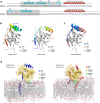
Fig 7. Convergence in the mechanisms of ACBD3 recruitment by enteroviruses and kobuviruses.
a, Distinct ACBD3-binding regions of enterovirus and kobuvirus 3A proteins. Sequences of the poliovirus-1 (member of enteroviruses) and aichivirus-1 (member of kobuviruses) 3A proteins are shown. Secondary structures present in the crystal structures of the ACBD3: 3A complexes (colored in light blue) and the hydrophobic alpha helices anchoring the 3A proteins to the membrane (colored in red) are indicated above the sequences. ACBD3-binding regions are shaded in grey. Myristoylated Gly1 of the aichivirus-1 3A protein is marked with an asterisk. b, Crystal structures of the ACBD3 GOLD domain in complex with the poliovirus (left) and aichivirus (right) 3A proteins. The protein backbones are shown in cartoon representation. The ACBD3 GOLD domain is depicted in grey, the viral 3A proteins in rainbow colors from blue (N terminus) to red (C terminus). c, Superposition of the crystal structures from (b). The ACBD3 GOLD domain is depicted in grey, the poliovirus 3A protein in blue, the aichivirus 3A protein in red. d, Molecular dynamics simulation-based models of the ACBD3 GOLD domain in complex with the poliovirus (left) and aichivirus (right) 3A proteins on the lipid bilayer. The ACBD3 GOLD domain is shown in cartoon representation with a semi-transparent surface and colored in gold except for the membrane binding site, which is colored in green. The poliovirus 3A protein is depicted in blue, the aichivirus 3A protein in red.

Similar articles

Cited by

References

    1. Tapparel C, Siegrist F, Petty TJ, Kaiser L. Picornavirus and enterovirus diversity with associated human diseases. Infect Genet Evol. 2013;14:282–93. 10.1016/j.meegid.2012.10.016 - DOI - PubMed
    1. Wessels E, Duijsings D, Niu T-K, Neumann S, Oorschot VM, de Lange F, et al. A viral protein that blocks Arf1-mediated COP-I assembly by inhibiting the guanine nucleotide exchange factor GBF1. Dev Cell. 2006;11:191–201. 10.1016/j.devcel.2006.06.005 - DOI - PubMed
    1. Greninger AL, Knudsen GM, Betegon M, Burlingame AL, DeRisi JL. The 3A protein from multiple picornaviruses utilizes the golgi adaptor protein ACBD3 to recruit PI4KIIIβ. J Virol. 2012;86:3605–16. 10.1128/JVI.06778-11 - DOI - PMC - PubMed
    1. Liao J, Guan Y, Chen W, Shi C, Yao D, Wang F, et al. ACBD3 is required for FAPP2 transferring glucosylceramide through maintaining the Golgi integrity. J Mol Cell Biol. 2018. 10.1093/jmcb/mjy030 - DOI - PMC - PubMed
    1. Sohda M, Misumi Y, Yamamoto A, Yano A, Nakamura N, Ikehara Y. Identification and characterization of a novel Golgi protein, GCP60, that interacts with the integral membrane protein giantin. J Biol Chem. 2001;276:45298–306. 10.1074/jbc.M108961200 - DOI - PubMed

Publication types

MeSH terms

Substances