. 2019 Aug 2;7(3):79.
doi: 10.3390/vaccines7030079.

Immunogenicity and Efficacy Evaluation of Subunit Astrovirus Vaccines

Affiliations

Immunogenicity and Efficacy Evaluation of Subunit Astrovirus Vaccines

Mehdi R M Bidokhti et al. Vaccines (Basel). .

Abstract

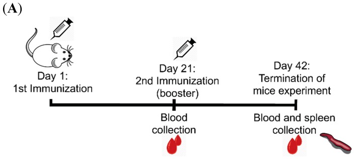
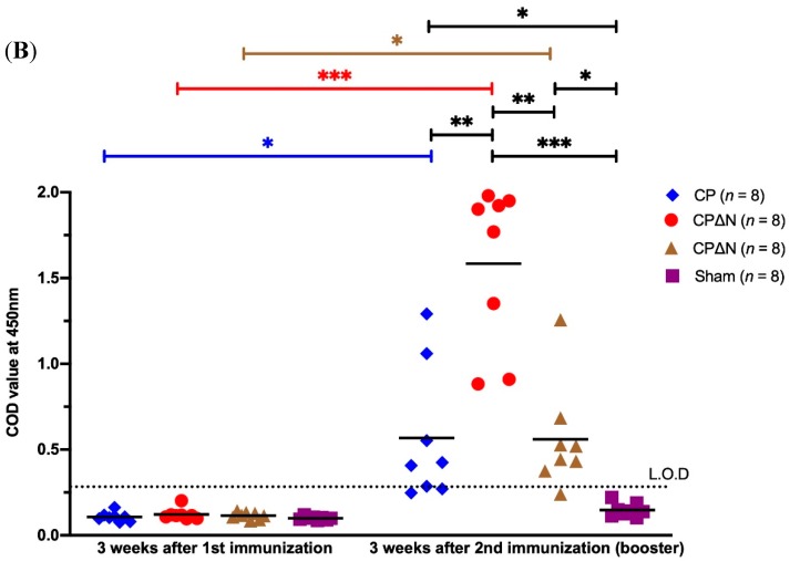
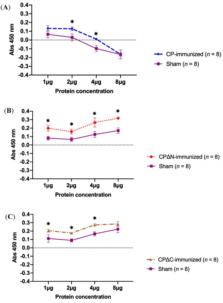
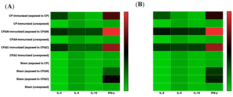
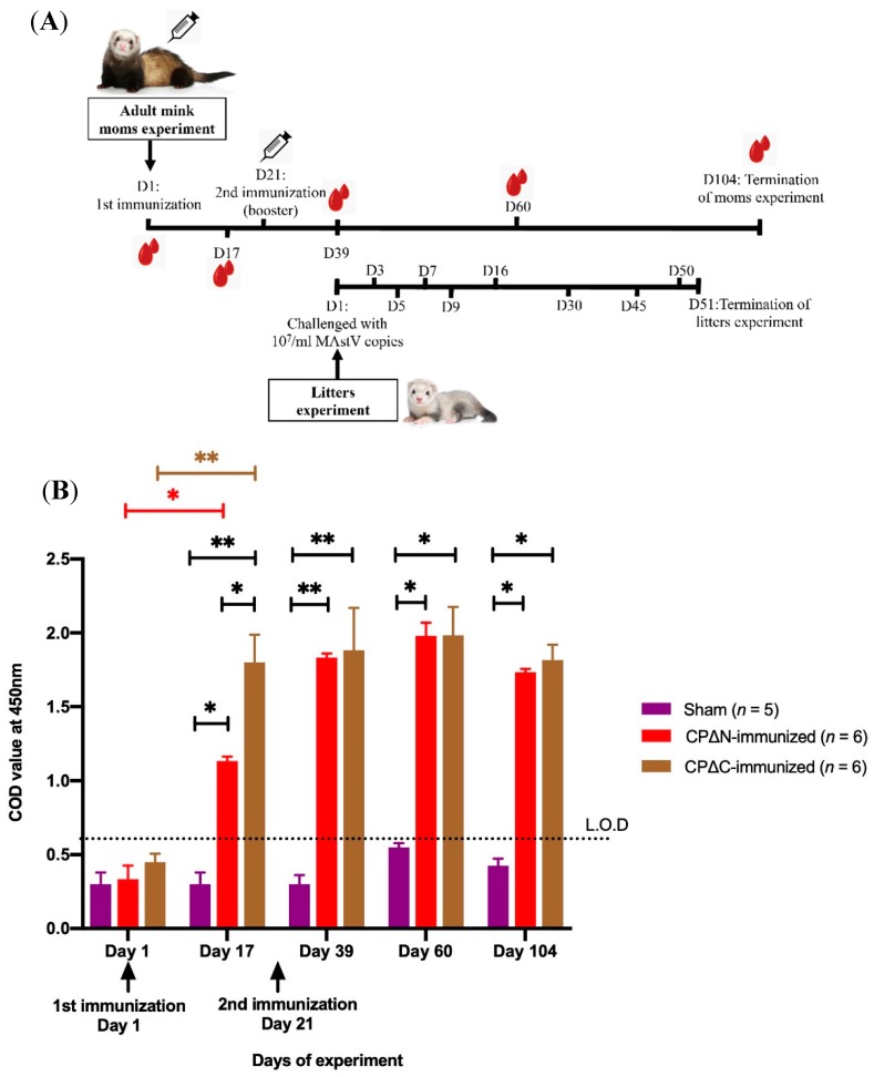
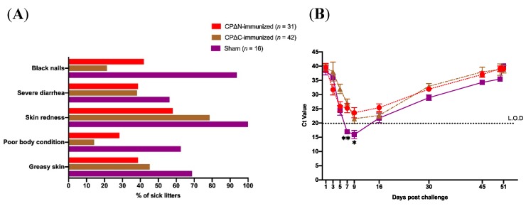
A full understanding of the immune response to astrovirus (AstV) infection is required to treat and control AstV-induced gastroenteritis. Relative contributions of each arm of the immune system in restricting AstV infection remain unknown. In this study, two novel subunit AstV vaccines derived from capsid protein (CP) of mink AstV (MAstV) such as CPΔN (spanning amino acids 161-775) and CPΔC (spanning amino acids 1-621) were evaluated. Their immunogenicity and cytokine production in mice, as well as protective efficacy in mink litters via maternal immunization, were studied. Truncated CPs induced higher levels of serum anti-CP antibodies than CP, with the highest level for CPΔN. No seronegativity was detected after booster immunization with either AstV CP truncates in both mice and mink. All mink moms stayed seropositive during the entire 104-day study. Furthermore, lymphoproliferation responses and Th1/Th2 cytokine induction of mice splenocytes ex vivo re-stimulated by truncated CPs were significantly higher than those by CP, with the highest level for CPΔN. Immunization of mink moms with truncated CPs could suppress virus shedding and clinical signs in their litters during a 51-day study after challenge with a heterogeneous MAstV strain. Collectively, AstV truncated CPs exhibit better parameters for protection than full-length CP.

Keywords: astrovirus; capsid protein; immunogenicity; infection; subunit vaccine.


Conflict of interest statement

The authors declare no conflict of interest. The founding sponsors had no role in the design of the study; in the collection, analyses, or interpretation of data; in the writing of the manuscript, and in the decision to publish the results.

Figures

Figure 1
Figure 1
Electron microscopy (EM) graph of mink astrovirus (MAstV), Danish strain DK7627 used for challenge experiment of litters. To prepare the infectious material for the experiment, fecal samples of a naturally-infected mink were examined by EM and small star-like particles of MAstV, 30 nm in diameter, were observed. Further analyses with a MAstV-specific PCR and sequencing also detected the genome of the MAstV Danish strain DK7627 in the sample. The number of RNA copies of MAstV in this infectious material measured by a quantitative PCR was 107/mL. This was later used for the challenge of litters. The white bar scale is 50 nm. The EM examination was performed at the Swedish Institute for Infectious Disease Control (SMI).
Figure 2
Figure 2
Schema of mice immunization study and indirect enzyme-linked immunosorbent assay (ELISA) titers of sera collected three weeks after the first and second immunization. (A) The MAstV vaccine candidates (5 µg/mouse) combined with an equal volume of AbISCO-100 adjuvant (10 µg/mouse) were injected subcutaneously to Naval Medical Research Institute (NMRI) mice (n = 8 per group, 4 weeks old) twice with a three-week interval. Mice injected with pDual-GC-vector-transfected cell lysate (5 µg/mouse) combined with adjuvant (10 µg/mouse) were analyzed as a sham group (n = 8). Spleen samples were collected at the end of the trial for ex vivo proliferation and cytokine analysis. (B) The serum samples of mice were collected three weeks after each immunization and tested with an indirect ELISA (Limit of detection (L.O.D) is the corrected optical density (COD) above 0.3). CP refers to the full-length capsid protein (spanning amino acids (aa) 1–775 of CP) of MAstV; CPΔN refers to an N-terminal truncated protein (spanning aa 161–775 of CP) of MAstV; CPΔC refers to a C-terminal truncated protein (spanning aa 1–621 of CP) of MAstV; Sham refers to the control group injected with pDual-GC-vector-transfected cell lysate. The mean value of each group (n = 8) of mice was calculated, the COD value of each mouse is illustrated in the dot plot. Asterisks indicate the level of significant difference between the mean value of either immunized or sham groups using two-way analysis of variance (ANOVA) with replication test (* p < 0.05, ** p < 0.01 and *** p < 0.001). Bars represent the mean value for each group.
Figure 2
Figure 2
Schema of mice immunization study and indirect enzyme-linked immunosorbent assay (ELISA) titers of sera collected three weeks after the first and second immunization. (A) The MAstV vaccine candidates (5 µg/mouse) combined with an equal volume of AbISCO-100 adjuvant (10 µg/mouse) were injected subcutaneously to Naval Medical Research Institute (NMRI) mice (n = 8 per group, 4 weeks old) twice with a three-week interval. Mice injected with pDual-GC-vector-transfected cell lysate (5 µg/mouse) combined with adjuvant (10 µg/mouse) were analyzed as a sham group (n = 8). Spleen samples were collected at the end of the trial for ex vivo proliferation and cytokine analysis. (B) The serum samples of mice were collected three weeks after each immunization and tested with an indirect ELISA (Limit of detection (L.O.D) is the corrected optical density (COD) above 0.3). CP refers to the full-length capsid protein (spanning amino acids (aa) 1–775 of CP) of MAstV; CPΔN refers to an N-terminal truncated protein (spanning aa 161–775 of CP) of MAstV; CPΔC refers to a C-terminal truncated protein (spanning aa 1–621 of CP) of MAstV; Sham refers to the control group injected with pDual-GC-vector-transfected cell lysate. The mean value of each group (n = 8) of mice was calculated, the COD value of each mouse is illustrated in the dot plot. Asterisks indicate the level of significant difference between the mean value of either immunized or sham groups using two-way analysis of variance (ANOVA) with replication test (* p < 0.05, ** p < 0.01 and *** p < 0.001). Bars represent the mean value for each group.
Figure 3
Figure 3
Proliferation assay of mice splenocytes after re-stimulation with different concentrations of the corresponding MAstV capsid proteins. The MAstV vaccine candidates (5 µg/mouse) combined with an equal volume of AbISCO-100 adjuvant (10 µg/mouse) were injected to NMRI mice (n = 8 per group, 4 weeks old) twice with a three-week interval. Mice injected with pDual-GC-vector-transfected cell lysate combined with adjuvant were also analyzed as sham (n = 8). Three weeks after the second immunization, the splenocytes were harvested from all mice groups: (A) CP-immunized (n = 8), (B) CP∆N-immunized (n = 8), and (C) CP∆C-immunized (n = 8). The splenocytes were then purified, cultivated ex vivo (2 × 10⁵ cells/well), and stimulated by exposing to different final concentrations (1 μg, 2 μg, 4 μg, and 8 μg) of the corresponding MAstV CPs as described in Materials and Methods; cell proliferation was measured in a WST-1 assay after 48 h of incubation. The splenocytes of the sham group (n = 8) were also independently exposed to each of the three MAstV CPs. The data are mean COD value readings of triplicate experiments. Asterisks indicate a significant difference at the given protein concentration between immunized and sham mice tested by two-way ANOVA with replication test (* p < 0.05). Error bars represent standard deviation (SD).
Figure 4
Figure 4
Heatmap of T-cell-mediated immune response to the corresponding MAstV CPs. The MAstV vaccine candidates (5 µg/mouse) combined with an equal volume of AbISCO-100 adjuvant (10 µg/mouse) were injected into NMRI mice (n = 8 per group, 4 weeks old) twice with a three-week interval. Mice injected with pDual-GC-vector-transfected cell lysate combined with adjuvant was also analyzed as a sham (n = 8). Three weeks after the second immunization, mice splenocytes were harvested from each group of mice: CP-immunized mice, CP∆N-immunized mice, and CP∆C-immunized mice. Mice injected with pDual-GC-vector-transfected cell lysate combined with adjuvant was also analyzed as a sham (n = 8). The splenocytes of four mice per group were then extracted, washed, and cultivated ex vivo (2 × 10⁵ cells/well) and stimulated by exposing to (A) 2 µg/mL and (B) 4 µg/mL of the corresponding MAstV CPs as described in Materials and Methods. The culture supernatants from stimulated splenocytes were collected and assessed for secreted cytokines IL-2, IL-5, IL-10, and IFN-γ by using a mouse Th1/Th2 Cytokine Cytometric 6-plex Array Bead kit (Invitrogen), Luminex® 100/200™ System and xPONENT® software (Luminex Corporation, Austin, TX, USA). The splenocytes of the sham group (n = 8) were also independently exposed to each of the three MAstV CPs. The data are mean COD readings of duplicate experiments, illustrated as log 10 values in the heatmap.
Figure 5
Figure 5
Schema of mink immunization and ELISA of the humoral immune response to mink astrovirus capsid protein (MAstV CP) vaccine candidates in adult mink moms. (A) The MAstV vaccine candidates (100 µg/animal) combined with an equal amount of Freund’s adjuvant were injected into female adult minks (n = 6 per group) twice with three weeks interval. The adult moms injected with pDual-GC-vector-transfected cell lysates combined with adjuvant were also analyzed as a sham (n = 5). On day (D) 1 after birth, the litters (n = 89) of these immunized and sham adult mink moms (n = 17) were then challenged orally with 107/mL MAstV copies. As a follow-up, the fecal samples of their newborn litters on test days 1, 3, 5, 7, 9, 16, 30, 45, 50, and 51 after birth were tested using real-time PCR for detecting astrovirus shedding status. The clinical signs of litters were also monitored and recorded by veterinarians every other day. (B) Serum samples of the adult moms were collected on test days 1, 17, 39, 60, and 104 and tested with an indirect ELISA for measuring maternal antibody production against MAstV (Limit of detection (L.O.D) is the corrected optical density (COD) above 0.6). CPΔN refers to an N-terminal truncated protein (spanning aa 161–775 of CP) of MAstV; CPΔC refers to a C-terminal truncated protein (spanning aa 1–621 of CP) of MAstV; Sham refers to the control group injected with pDual-GC-vector-transfected cell lysate. Asterisks indicate significant differences at the given protein concentration between various immunized and sham adult mink moms tested by a two-way ANOVA with replication test (* p < 0.01, ** p < 0.001). Error bars represent SD.
Figure 6
Figure 6
Graphs of the results of the real-time polymerase chain reaction (PCR) and clinical signs observed in the experiment of litter minks. After birth, the one-day newborn litters (n = 89) of immunized and sham adult moms (n = 17) were challenged with 107/mL MAstV copies. As a follow-up, the fecal samples (n = 414) of their newborn litters on test days 1, 3, 5, 7, 9, 16, 30, 45, 50, and 51 post-challenge were tested using a real-time PCR for detecting AstV shedding status. The clinical signs of litters were also monitored and recorded by the author veterinarians every other day. (A) The ratios of litters showing various clinical signs to total group-related litters were calculated and illustrated as a percentage (%) in the graph. (B) The mean cycle threshold (Ct) value of real-time PCR results of sampled litters on each sampling occasion during challenge experiment (51 days) was calculated and is illustrated in the graph. Asterisks show a significant decline of virus shedding among litters of either both CP∆N- and CP∆C- (**) or just CP∆N- (*) immunized adult mom groups compared to those of the sham group using the Log-rank test (p < 0.05). Bars indicate the standard deviation (SD).

Similar articles

Cited by

References

    1. Holmgren J., Svennerholm A.M. Vaccines against mucosal infections. Curr. Opin. Immunol. 2012;24:343–353. doi: 10.1016/j.coi.2012.03.014. - DOI - PubMed
    1. Plotkin S.A. Vaccines, vaccination, and vaccinology. J. Infect. Dis. 2003;187:1349–1359. doi: 10.1086/374419. - DOI - PubMed
    1. Suschak J.J., Williams J.A., Schmaljohn C.S. Advancements in DNA vaccine vectors, non-mechanical delivery methods, and molecular adjuvants to increase immunogenicity. Hum. Vaccines Immunother. 2017;13:2837–2848. doi: 10.1080/21645515.2017.1330236. - DOI - PMC - PubMed
    1. Tan M., Jiang X. Recent advancements in combination subunit vaccine development. Hum. Vaccines Immunother. 2017;13:180–185. doi: 10.1080/21645515.2016.1229719. - DOI - PMC - PubMed
    1. Humphreys I.R., Sebastian S. Novel viral vectors in infectious diseases. Immunology. 2018;153:1–9. doi: 10.1111/imm.12829. - DOI - PMC - PubMed

LinkOut - more resources