. 2019 Aug 2;9(8):336.
doi: 10.3390/biom9080336.

Multiple Modulation of Acid-Sensing Ion Channel 1a by the Alkaloid Daurisoline

Affiliations

Multiple Modulation of Acid-Sensing Ion Channel 1a by the Alkaloid Daurisoline

Dmitry I Osmakov et al. Biomolecules. .

Abstract

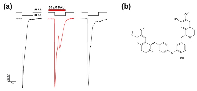
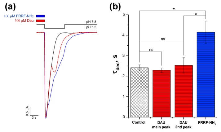
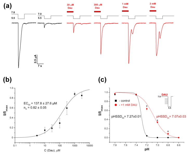
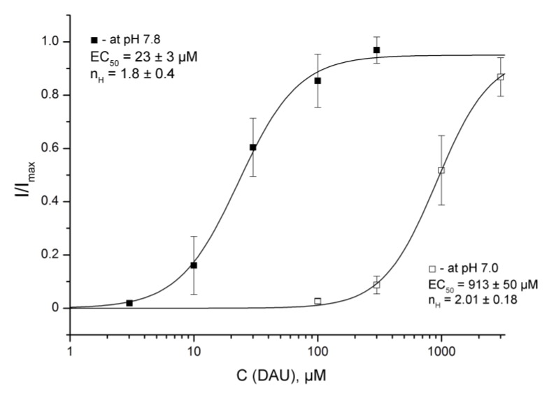
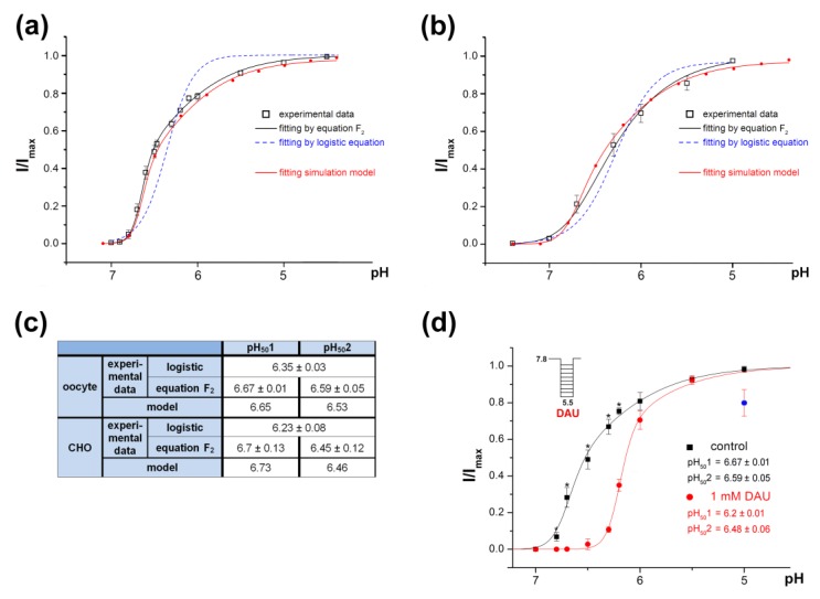
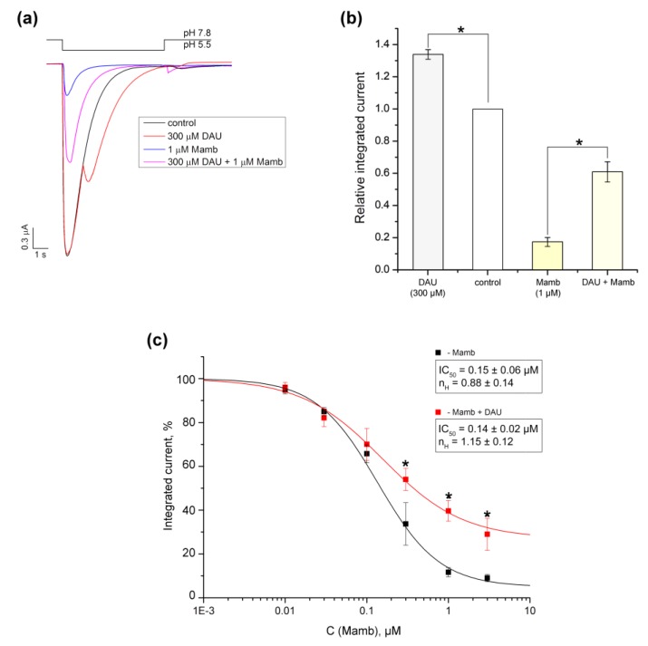
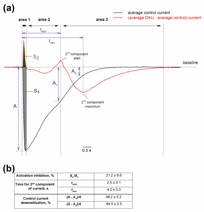
Acid-sensing ion channels (ASICs) are proton-gated sodium-selective channels that are expressed in the peripheral and central nervous systems. ASIC1a is one of the most intensively studied isoforms due to its importance and wide representation in organisms, but it is still largely unexplored as a target for therapy. In this study, we demonstrated response of the ASIC1a to acidification in the presence of the daurisoline (DAU) ligand. DAU alone did not activate the channel, but in combination with protons, it produced the second peak component of the ASIC1a current. This second peak differs from the sustained component (which is induced by RF-amide peptides), as the second (DAU-induced) peak is completely desensitized, with the same kinetics as the main peak. The co-application of DAU and mambalgin-2 indicated that their binding sites do not overlap. Additionally, we found an asymmetry in the pH activation curve of the channel, which was well-described by a mathematical model based on the multiplied probabilities of protons binding with a pool of high-cooperative sites and a single proton binding with a non-cooperative site. In this model, DAU targeted the pool of high-cooperative sites and, when applied with protons, acted as an inhibitor of ASIC1a activation. Moreover, DAU's occupation of the same binding site most probably reverses the channel from steady-state desensitization in the pH 6.9-7.3 range. DAU features disclose new opportunities in studies of ASIC structure and function.

Keywords: ASIC1a channels; channel desensitization; daurisoline; potentiator.


Conflict of interest statement

The authors declare no conflict of interest.

Figures

Figure 1
Figure 1
Effect of daurisoline (DAU) application on a rat ASIC1a channel and its structure. (a) Potentiating effect of DAU (30 μM) on ASIC1a channel activation by fast external solution acidification from pH 7.8 to 5.5. The whole-cell currents shown were obtained from the same oocyte. (b) Chemical formula of DAU.
Figure 2
Figure 2
Concentration-dependent activation of the DAU-induced second component of the ASIC1a current. (a) Representative whole-cell traces for currents evoked by a fast pH 5.5 stimulus in the control (black) and in the channels that were pre-incubated for 15 s with DAU at various concentrations (red). (b) The dose–response curve fitted to the logistic equation for the second component of the ASIC1a current. I/Imax is the amplitude of the second component of the current at a conditioning pH of 7.8, normalized to the maximum amplitude (Imax) calculated for each oocyte by individual fitting. Data are presented as mean ± SEM (n = 6).
Figure 3
Figure 3
Comparison of the effects that DAU and the RF-amide peptide have on ASIC1a current. (a) Representative traces of pH 5.5-evoked currents of ASIC1a alone (black trace) and of ASIC1a with a 15 s pre-incubation of 300 μM DAU (red trace) or 100 μM FRRF-NH2 peptide (blue trace). (b) Effects of 300 μM DAU and 100 μM FRRF-NH2 peptide on the time of exponential decay (τdec) for the main peak (both control and with the peptide) and for the second (DAU) component of the current (DAU). Time τdec was calculated as the interval between the maximum of the current amplitude and the point having 1/e times reduced amplitude to this maximum. The significance of data was proved by statistical analysis of variance followed by a Tukey’s test ANOVA. Results are presented as mean ± SEM (n = 5–12); ns—non significantly difference; * P < 0.05.
Figure 4
Figure 4
DAU-induced inhibition of steady-state desensitization (SSD) of rat ASIC1a. (a) Whole-cell currents obtained from the channels held at the conditioning pHs of 7.8 and 7.0 in response to a pH-5.5 stimulus, with (red) or without (black) a pre-incubation for 15 s with DAU at various concentrations. (b) The dose–response curve fitted to the logistic equation for peak ASIC1a amplitudes at various concentrations of pre-incubated DAU. I/Imax is the main peak’s amplitude at a conditioning pH of 7.0, normalized to the maximum amplitude (Imax), which we calculated for each oocyte via individual fitting. Data are presented as mean ± SEM (n = 6). (c) pH dependence of the channel’s SSD without (black curve) and with (red curve) pre-incubation of 1 mM DAU. ASIC1a held at various conditioning pHs was activated by same pH 5.5 stimulus. The peak current amplitudes are plotted as a function of pH and fitted to the logistic equation. Data are presented as mean ± SEM (n = 5); * P < 0.05.
Figure 5
Figure 5
DAU-induced development of the second peak at conditioning pHs of 7.8 and 7.0. Dose–response curves for the second peak by DAU pre-incubation at the holding pHs of 7.8 and 7.0 were plotted for same pH 5.5 activating stimulus. I/Imax is the second peak’s amplitude at a conditioning pH of 7.8 or 7.0, normalized to the maximum amplitude (Imax), which we calculated for each oocyte via individual fitting. Data are presented as mean ± SEM (n = 6).
Figure 6
Figure 6
The peculiarity of rat ASIC1a activation by protons. (a) pH dependence of activation of ASIC1a channels expressed in X. laevis oocytes fitted to the logistic equation F1 (dotted line) and to equation F2 (black solid line). Redline reflects pH dependence of activation of model 3–10 corresponding to activation of the channel at the binding of 10 protons with coefficients of cooperativity of p = r = 0.8 and q = s = 0.65, respectively, fitted to equation F2. (b) pH dependence of activation of the channels expressed in CHO cells fitted to the logistic equation F1 (dashed line) and to equation F2 (black solid line). The data were re-plotted from Figure 1k in [38]. Redline reflects pH dependence of activation of model 3–10 corresponding to activation of the channel at the binding of 10 protons with coefficients of cooperativity of p = r = 0.79 and q = s = 0.6, respectively, fitted to equation F2. (c) Calculated parameters of pH50 for non-discriminated sites, pH501 for “high-cooperative” sites and pH502 for “non-cooperative” site are shown as a result of experimental data mathematical processing. (d) pH dependence of ASIC1a activation by protons (black line) and by the combination of protons and 1 mM DAU (red line). ASIC1a held at pH 7.8 was activated by various acidic pH stimuli. I/Imax is the amplitude of the peak current evoked by acidic pH stimuli and normalized to the maximum amplitude (Imax), which we calculated for each cell via individual fitting. Data are presented as mean ± SEM (n = 7); * P < 0.05.
Figure 7
Figure 7
Comparison of the effects that DAU and Mamb-2 have on ASIC1a. (a) Representative traces of pH 5.5-evoked currents of ASIC1a alone (black trace) and of ASIC1a with a 15 s pre-incubation of 300 μM DAU (red trace), 1 μM mambalgine-2 peptide (Mamb-2) (blue trace), or a mixture of 300 μM DAU and 1 μM Mamb-2 (magenta trace). (b) Integrated currents measured for ASIC1a (alone and in the presence of each of the following: 300 μM DAU, 1 μM Mamb-2, and a mixture of 300 μM DAU and 1 μMMamb-2) generated by dropping the pH from 7.8 to 5.5. Data are presented as mean ± SEM; n = 5. *P < 0.05, significantly different from the integrated current at pH 7.8; Kruskal–Wallis ANOVA. (c) Dose–response ASIC1a inhibitory curves for Mamb-2 alone (black line) and for Mamb-2 in concurrence with 300 μM DAU (red line). Data were fitted by the logistic equation. The calculated permanent current for Mamb-2 was 5.3 ± 2.3%, and the maximal integrated current increased to 26.7 ± 7.8% in the presence of DAU. Data are presented as mean ± SEM (n = 5); * P < 0.05.
Scheme 1
Scheme 1
Model 3–10 with ten consecutive H+ binding steps.
Figure 8
Figure 8
Dual effect of pre-incubated DAU on ASIC1a channel. (a) Average control current (n = 7; black trace) and current obtained by subtracting the average control current from the average current induced by 300 µM DAU (n = 7; red trace). The black arrows indicate the boundary of the three conditional zones of the DAU action: Area 1 (inhibitory), area 2 (neutral), and area 3 (potentiating). For area 1, we calculated the integral control current S1 (grey) and the integral inhibited current S2 (green). A is the maximal amplitude of the control current; A1 and A2 are the amplitudes of the control current at time tstart (the start of second peak’s development) and tmax (the second peak’s maximum), respectively. (b) Calculated parameters of the DAU effect, presented as mean ± SEM (n = 7). The inhibition of the channel activation as a ratio of area S2 to area S1 is expressed as a delay in the of channel’s opening kinetics without a change in the peak amplitude (A). We estimated the percentage by which the main current is desensitized at tstart and tmax using the ratio of the difference between amplitudes (A minus A1 and A2, respectively) to amplitude A.

References

    1. Waldmann R., Champigny G., Bassilana F., Heurteaux C., Lazdunski M. A proton-gated cation channel involved in acid-sensing. Nature. 1997;386:173–177. doi: 10.1038/386173a0. - DOI - PubMed
    1. Jasti J., Furukawa H., Gonzales E.B., Gouaux E. Structure of acid-sensing ion channel 1 at 1.9 Å resolution and low pH. Nature. 2007;449:316–323. doi: 10.1038/nature06163. - DOI - PubMed
    1. Bartoi T., Augustinowski K., Polleichtner G., Grunder S., Ulbrich M.H. Acid-sensing ion channel (ASIC) 1a/2a heteromers have a flexible 2:1/1:2 stoichiometry. Proc. Natl. Acad. Sci. USA. 2014;111:8281–8286. doi: 10.1073/pnas.1324060111. - DOI - PMC - PubMed
    1. Deval E., Gasull X., Noël J., Salinas M., Baron A., Diochot S., Lingueglia E. Acid-Sensing Ion Channels (ASICs): Pharmacology and implication in pain. Pharmacol. Ther. 2010;128:549–558. doi: 10.1016/j.pharmthera.2010.08.006. - DOI - PubMed
    1. Wemmie J.A., Taugher R.J., Kreple C.J. Acid-sensing ion channels in pain and disease. Nat. Rev. Neurosci. 2013;14:461–471. doi: 10.1038/nrn3529. - DOI - PMC - PubMed

Publication types