Review
. 2019 Aug 2;24(15):2826.
doi: 10.3390/molecules24152826.

Real-Time 3D Single Particle Tracking: Towards Active Feedback Single Molecule Spectroscopy in Live Cells

Affiliations
Review

Real-Time 3D Single Particle Tracking: Towards Active Feedback Single Molecule Spectroscopy in Live Cells

Shangguo Hou et al. Molecules. .

Abstract

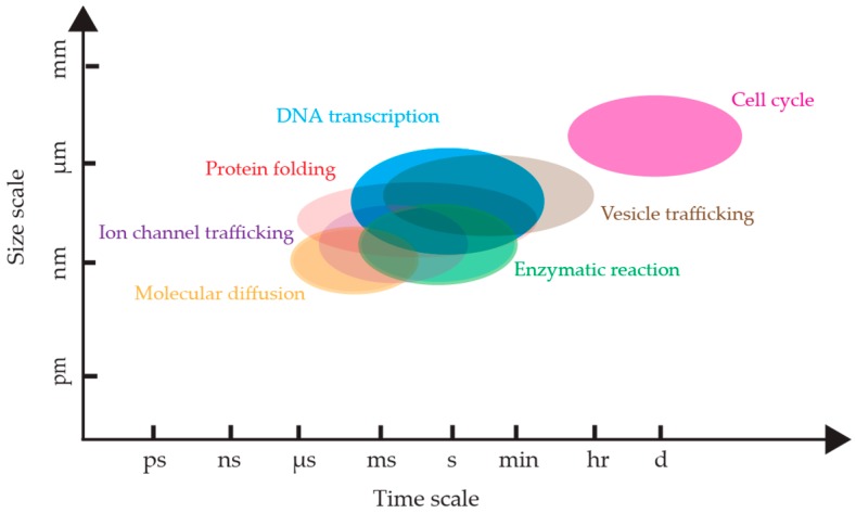
Single molecule fluorescence spectroscopy has been largely implemented using methods which require tethering of molecules to a substrate in order to make high temporal resolution measurements. However, the act of tethering a molecule requires that the molecule be removed from its environment. This is especially perturbative when measuring biomolecules such as enzymes, which may rely on the non-equilibrium and crowded cellular environment for normal function. A method which may be able to un-tether single molecule fluorescence spectroscopy is real-time 3D single particle tracking (RT-3D-SPT). RT-3D-SPT uses active feedback to effectively lock-on to freely diffusing particles so they can be measured continuously with up to photon-limited temporal resolution over large axial ranges. This review gives an overview of the various active feedback 3D single particle tracking methods, highlighting specialized detection and excitation schemes which enable high-speed real-time tracking. Furthermore, the combination of these active feedback methods with simultaneous live-cell imaging is discussed. Finally, the successes in real-time 3D single molecule tracking (RT-3D-SMT) thus far and the roadmap going forward for this promising family of techniques are discussed.

Keywords: active feedback tracking; real-time 3D single particle tracking; single molecule spectroscopy.


Conflict of interest statement

The authors declare no conflict of interest.

Figures

Figure 1
Figure 1
The temporal and spatial scales of biological processes. The time windows and size windows are approximate, and each process has its own situation-dependent scale. References: DNA transcription rate [29], protein folding [30,31,32], molecular diffusion [33,34,35], cell cycle [36], vesicle trafficking [37,38,39,40], ion channel trafficking [41,42,43], enzymatic reactions [3,44].
Figure 2
Figure 2
(A) Top row: The solution-phase single molecule observation limits measurement time to the diffusion time in the laser focus. Middle row: Tethering to the coverslip extends the observation time but precludes translation to live-cell studies. Bottom row: Real-time 3D single molecule tracking spectroscopy potentially enables continuous observation of freely diffusing proteins in the cellular interior. (B) An example of a simulated lifetime of a single fluorophore labelled protein diffusing through a confocal volume, emitting on the order of 10 photons, yielding a large experimental error.
Figure 3
Figure 3
Overview of active feedback tracking methods covered in this review.
Figure 4
Figure 4
Tetrahedral detection active feedback 3D tracking. (A) Schematic of experimental setup. BS: beamsplitter; SPAD: single-photon counting avalanche photodiode. (B,D) Schematic of the sample volume viewed along Z (B) and Y (D) axis, in which the four balls represent the sample volumes of four SPC-APDs. (C,E) Total fluorescent intensity in the Z = 0 plane (C) and Y = 0 plane (E). Reprinted from reference [67], with the permission of AIP Publishing. (F) 3D trajectory of a single quantum dot labeled high-affinity immunoglobin E receptor (IgE-FcεR) in a live cell. The rainbow color from red to blue indicates time. (G) Image of cell and the actively tracked receptor (white arrow). (H) Fluorescence intensity of the quantum dot probe as a function of time. (I) Photon pair correlation measurement shows fluorescence photon antibunching, indicating a single quantum emitter. Reprinted with permission from reference [70]. Copyright 2010 American Chemical Society.
Figure 5
Figure 5
Multi-detector active feedback 3D tracking. (A) Schematic of dark-field multi-detector active feedback tracking system developed by Cang et al. which utilized a quadrant photodiode. Reprinted from reference [72], with the permission of AIP Publishing. (B) Schematic of multi-detector setup used for fluorescent nanoparticle tracking. (C) Top-down view of a polystyrene-quantum dot-peptide nanoparticle (PS-QD-peptide) landing on a membrane protrusion of a live cell. (D,E) High-resolution view of trajectory in (C) with different angles. (F) Dynamics heat map of the trajectory, showing regions of varying particle diffusivity depending on its location on the nanoscale protrusion (C). (G) Overlaid image of two-photon live cell image and high-resolution 2D trajectory. (H) High-resolution 3D trajectory of a PS-QD-peptide as it traces out hemispherically-capped protrusions on the live cell membrane. (I) Diffusional states as a function of time analyzed by change-point diffusion analysis. (J) Dynamics heat map of trajectory in (H). Adapted from reference [76] with permission of the author.
Figure 6
Figure 6
Concept of orbital tracking. The laser (green) is scanned laterally in a circular pattern around a particle (orange dot). (A) When a particle deviates from the center of the orbital scan, the fluorescence intensity (I0, red curve in the low panel) of the particle is modulated at the same frequency as the laser scan. The phase (ϕ0) and intensity (I1) are extracted via FFT or lock-in amplifier and used to calculate the position of the particle within the orbit. The calculated position is used to relocate the particle so that the modulation of intensity is minimized. (B) When a particle is located in the center of the orbital scan, the fluorescence intensity of the particle remains stable with time.
Figure 7
Figure 7
Orbital tracking with biplane detection. (A) Schematic of particle position measurement. The laser focus is scanned in a circle around the tracked particle while the two detection channels monitor the intensity at two axial planes (right panel). The X and Y positions of the particle are calculated from the modulation amplitude and phase of the fluorescence signal. The Z position is calculated by the difference of the signal between the two detection channels. Reprinted from reference [84]. Copyright 2009 Wiley-VCH. (B) 3D orbital tracking of single-walled carbon nanotubes (SWNT) in live cells. The image shows overlaid bright field image of Hela cell (outlined in yellow) with 18 SWNT trajectories. Adapted from reference [85]. Copyright 2012 American Chemical Society.
Figure 8
Figure 8
(A) Schematic of orbital 3D tracking with high-speed laser modulation using acousto-optics. Two laser beams with rotation frequency ωxy are focused at two different depths in the sample separated by ~1 µm. The excitation power is alternated between these two laser beams with a frequency of ωz. The magnitude of the ωz frequency component in the fluorescence intensity is proportional to the particle’s distance from z = 0. Adapted from reference [88]. Copyright 2007 American Chemical Society. (B) Schematic of 3D orbital tracking using an ETL. A complete 3D tracking period is comprised of two orbits above the particle and two orbits below the particle. (C) Comparison of the response time of piezo stage versus ETL for an orbital scanning period of 8.192 ms, with the ETL showing a much faster response. Adapted from reference [83]. Copyright 2015 Optical Society of America.
Figure 9
Figure 9
(A) Schematic of tetrahedral excitation tracking setup, where four laser diode beams are combined by three beamsplitters and focused onto the sample in a tetrahedral pattern. (inset) Washers inserted between the fiber coupler (FC) and collimator (Col) are used to arrange the four laser foci after the objective. Adapted from reference [91]. Copyright 2015 Optical Society of America. (B) Schematic of TSUNAMI. A pulsed laser is split by two beamsplitters to generate four laser beams. The temporal separation between four beams is achieved by physical delay. The arrangement of the four foci after the objective is controlled by mirrors in the excitation beam path. (c) Temporal separation of the four excitation beams measured by the fluorescence of an emitter in the center of the detection volume. (D) Schematic of the tetrahedral PSF. (E) Scanning mage of 100 nm fluorescent beads in different depth. Scale bar: 2 μm. Adapted from reference [92] under Creative Commons Attribution 4.0 International License.
Figure 10
Figure 10
(A) Schematic of 3D-DyPLoT. A 2D-EOD is used to deflect the laser in X and Y in a knight’s tour pattern (20 μs per spot) while a TAG lens dynamically modulates the focus at ~ 70 kHz. (B) Fixed 190 nm fluorescent particle is driven along a predetermined pattern by a micro-stage and tracked by 3D-DyPLoT. (CF) 3D-DyPLoT is used to track silica-coated giant nonblinking quantum dots (gQD) in water. (C) 3D trajectory. (D) Fluorescence intensity of gQD as a function of time. (E) MSD analysis of a free diffusing gQD. (F) Histogram of diffusion coefficient and calculated diameter of gQD showing a mean hydrodynamic diameter of 38 nm. (inset) TEM of silica-coated gQD. Adapted from reference [93]. Copyright 2017 Optical Society of America.
Figure 11
Figure 11
(A) Schematic of image-based biplane active feedback 3D tracking. One part of fluorescence is transmitted through a 50:50 beamsplitter and imaged onto the EMCCD camera while the other part of fluorescence is reflected by beamsplitter and imaged on a different side of the EMCCD. The reflected fluorescence has a longer path so that two axial planes can be monitored synchronously. The piezo mirror and Z piezo stage are used to move the laser focus in 3D. Adapted from reference [100]. Copyright 2010 American Chemical Society. (B) Schematic of astigmatic imaging active feedback 3D tracking. A high NA objective is used for light sheet illumination and the 3D information of the particle is encoded in the astigmatic image. The image is analyzed and a feedback control is applied to the Z piezo stage to maintain the particle in focus. Adapted from reference [101] under Creative Commons CC BY Licence.
Figure 12
Figure 12
Prior volumetric imaging combined with TSUNAMI-based tracking was used to acquire a volumetric image of the entire 100 µm diameter tumor spheroid around an internalizing particle. (A) 3D isocontour reconstruction of tumor spheroid with red plasma membrane and blue nucleus. Green section at 50 µm shows plane of EGFR internalization trajectory. (B) Isocontour model of the green slice in (A). Inset shows zoomed in view highlighting spheroid boundary and the placement of the trajectory as it transports into the spheroid. (C) Zoom in (B). (D) Isolated trajectory. Adapted from reference [92] under Creative Commons Attribution 4.0 International License.
Figure 13
Figure 13
The addition of simultaneous widefield imaging to orbital tracking allowed trajectories (blue) of artificial viruses (red) to be placed in the environmental context of their interactions with the cytoskeleton as visualized with eGFP-labeled tubulin (green). (A) Trajectory of an artificial virus shown in 3D space with 2D projections across each axis shown in gray. (B) Two frames in a time series of images with trajectory overlaid. In the left frame, the particle is moving laterally not because the virus itself is changing microtubules, but because the microtubule itself was moving. This observation could not have been confirmed without simultaneously acquired imaging. Reprinted from reference [84]. Copyright 2009 Wiley-VCH.
Figure 14
Figure 14
Tetrahedral detection tracking coupled with simultaneously acquired spinning disk confocal images. (A) Localization of QD-labeled probe within the cellular structured using LED illumination requiring 300 ms exposure time. (B) Laser excitation reduced needed exposure time to 40 ms and provides a much higher contrast image than (A). Reprinted from reference [102], with the permission of AIP Publishing.
Figure 15
Figure 15
3D Multi-Resolution Microscopy combines RT-3D-SPT with simultaneously acquired 2P-LSM images. (A) Isolated trajectory of QD-labeled nanoparticle probe. (BD) Corresponding 2P-LSM sections acquired at depths indicated in circled portions of trajectory in (A). (E) Overlay of co-registered trajectory with 3D reconstruction of interpolated 2P-LSM sections gives cellular context to trajectory motion. Nuclear regions are highlighted in red and cell membrane surfaces are green. (F) 2P-LSM maximum intensity projection of internalized QD-labeled probe in a NIH-3T3 fibroblast cell. Arrow indicates position of tracked particle. (G) Ellipsoidal trajectory of macropinosomal membrane-bound probe showing multiple curved surfaces due to macropinosomal motion. (H) Structural tracing of the trajectory shown in (E). Arrow shows center of mass motion during trajectory. Adapted from reference [76] with permission of the author.
Figure 16
Figure 16
RT-3D-SMT using the tetrahedral detection method reveals the protein oligomerization states of Azami Green oligomers mAG, dAG, and tAG in 92% glycerol solution. Reprinted with permission from reference [104]. Copyright 2012 American Chemical Society.
Figure 17
Figure 17
Tetrahedral detection tracking of intramolecular FRET within double-stranded DNA in 90% glycerol-water solution. (A) X, Y, Z position and fluorescent intensity of AF488 (Alexa Fluor 488; ex: 490 nm; em: 525 nm)-DNA-AF594 (Alexa Fluor 4594; ex: 590 nm; em: 617 nm) molecule. (B) Fluorescence intensity and FRET efficiency as a function of time for the trajectory shown in (A). (C) Distribution of FRET efficiency of AF488-DNA molecules (control sample). (D) Distribution of FRET efficiency of AF488-DNA-AF594 molecules. Reprinted with permission from reference [71]. Copyright 2018 American Chemical Society. (E–I) Measurement of the hybridization kinetics of freely diffusing ssDNA molecules in solution by tetrahedral detection 3D tracking. (E) Scheme of the donor-quencher system for the DNA hybridization kinetics measurement. Red ball denotes ATTO633 (ex: 630 nm; em: 651 nm) reporter and grey ball denotes Iowa Black quencher. (F) Fluorescence lifetimes of ATTO633 with different donor-quencher separation distances. (G) Lifetime measurement in 3D tracking of ssDNA in 70 wt% glycerol solution. The fluorescence lifetime switching indicates transient annealing and melting events. (H) Lifetime histogram built from (G) showing two states. (I) Apparent annealing rate (kon) and melting rate (koff) as a function of quencher concentration shows the annealing rate is quencher concentration dependent. Adapted from reference [105]. Copyright 2017 the Royal Society of Chemistry.

Similar articles

Cited by

References

    1. Moerner W.E., Orrit M. Illuminating Single Molecules in Condensed Matter. Science. 1999;283:1670–1676. doi: 10.1126/science.283.5408.1670. - DOI - PubMed
    1. Ha T., Enderle T., Ogletree D.F., Chemla D.S., Selvin P.R., Weiss S. Probing the interaction between two single molecules: Fluorescence resonance energy transfer between a single donor and a single acceptor. Proc. Natl. Acad. Sci. 1996;93:6264. doi: 10.1073/pnas.93.13.6264. - DOI - PMC - PubMed
    1. Lu H.P., Xun L., Xie X.S. Single-Molecule Enzymatic Dynamics. Science. 1998;282:1877–1882. doi: 10.1126/science.282.5395.1877. - DOI - PubMed
    1. Shashkova S., Leake M.C. Single-molecule fluorescence microscopy review: Shedding new light on old problems. Biosci. Rep. 2017;37:BSR20170031. doi: 10.1042/BSR20170031. - DOI - PMC - PubMed
    1. Stracy M., Uphoff S., de Leon F.G., Kapanidis A.N. In vivo single-molecule imaging of bacterial DNA replication, transcription, and repair. FEBS Lett. 2014;588:3585–3594. doi: 10.1016/j.febslet.2014.05.026. - DOI - PubMed

LinkOut - more resources