Review
. 2019 Aug 2;8(8):818.
doi: 10.3390/cells8080818.

Cartilage and Bone Destruction in Arthritis: Pathogenesis and Treatment Strategy: A Literature Review

Affiliations
Review

Cartilage and Bone Destruction in Arthritis: Pathogenesis and Treatment Strategy: A Literature Review

Daisuke Tateiwa et al. Cells. .

Abstract

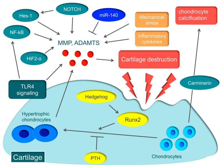
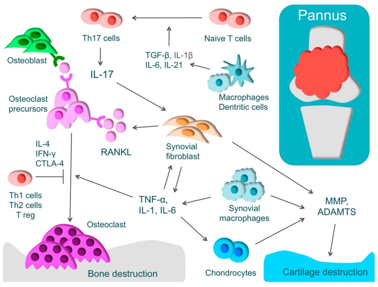
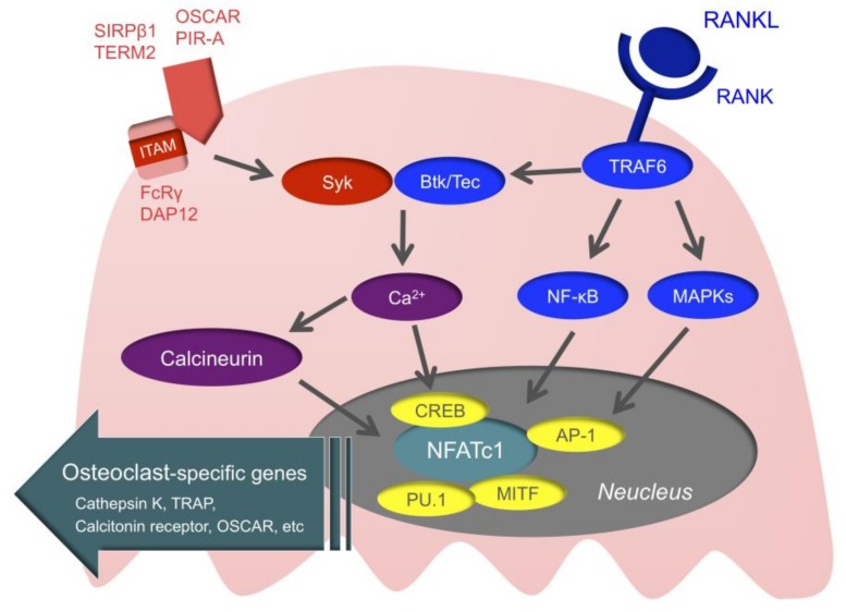
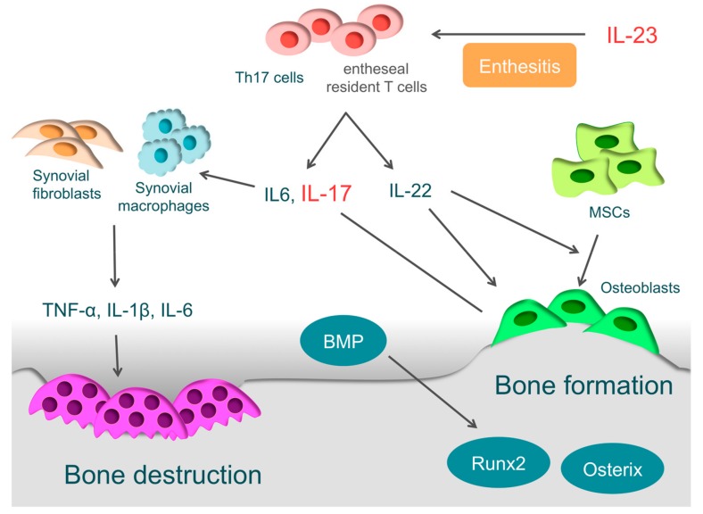
Arthritis is inflammation of the joints accompanied by osteochondral destruction. It can take many forms, including osteoarthritis, rheumatoid arthritis, and psoriatic arthritis. These diseases share one commonality-osteochondral destruction based on inflammation. The background includes a close interaction between osseous tissues and immune cells through various inflammatory cytokines. However, the tissues and cytokines that play major roles are different in each disease, and as a result, the mechanism of osteochondral destruction also differs. In recent years, there have been many findings regarding not only extracellular signaling pathways but also intracellular signaling pathways. In particular, we anticipate that the intracellular signals of osteoclasts, which play a central role in bone destruction, will become novel therapeutic targets. In this review, we have summarized the pathology of arthritis and the latest findings on the mechanism of osteochondral destruction, as well as present and future therapeutic strategies for these targets.

Keywords: arthritis; cartilage and bone destruction; osteoarthritis; osteoclast; psoriatic arthritis; rheumatoid arthritis.


Conflict of interest statement

The authors declare no conflict of interest.

Figures

Figure 1
Figure 1
Various molecular mechanisms associated with osteoarthritis (OA) Hypertrophic chondrocytes are found in abundance in the early stage of articular cartilage destruction, from which matrix metalloproteinase (MMP) and a disintegrin and metalloproteinase with thrombospondin motifs (ADAMTS) are secreted, triggering OA. The essential transcription factor in the hypertrophy of chondrocytes is runt-related transcription factor 2 (Runx2) that is regulated by Hedgehog. On the other hand, parathyroid hormone (PTH) suppresses the hypertrophy of chondrocytes. Hypoxia-inducible factor-2α (HIF-2α), nuclear factor-kappa B (NF-κB), Notch, Hes1, and toll-like receptor 4 (TLR4) signaling promote the secretion of various degrading enzymes, including MMP and ADAMTS, while miR-140 protects cartilages by suppressing the expression of ADAMTS5. Carminerin is a cartilage-specific protein involved in chondrocyte calcification.
Figure 2
Figure 2
The pathology of rheumatoid arthritis (RA) and the mechanism of cartilage and bone destruction. RA is characterized by proliferative synovium (pannus) and an excessive immune response of T-cells. Pannus comprises T-cells, synovial fibroblasts, and macrophages that produce inflammatory cytokines, such as tumor necrosis factor (TNF)-α, interleukin-1 (IL-1), IL-6, and IL-17. These inflammatory cytokines activate osteoclasts, leading to bone destruction. T-helper (Th) cells have subsets such as Th1, Th2, and Th17 cells. Naive T-cells differentiate into Th17 cells through IL-1β, IL-6, IL-21, and transforming growth factor-β (TGF-β). Th17 cells produce IL-17 that activates inflammation by acting on various immune cells and activates osteoclasts by inducing the receptor activator of nuclear factor kappa B ligand (RANKL) in synovial fibroblasts. IFN-γ, IL-4, and cytotoxic T-lymphocyte-associated protein 4 (CTLA-4), produced by Th1, Th2, and Treg, respectively, regulate osteoclast differentiation. Cartilage destruction is caused by matrix metalloproteinase (MMP) and a disintegrin and metalloproteinase with thrombospondin motifs (ADAMTS) produced by chondrocytes, synovial fibroblasts, and synovial macrophages.
Figure 3
Figure 3
Intracellular signals of osteoclasts. When the receptor activator of nuclear factor kappa B (RANK) is stimulated, TNF receptor-associated factor 6 (TRAF6) is recruited to RANK. Then, mitogen-activated kinases (MAPKs), nuclear factor-kappa B (NF-κB), and activator protein1 (AP-1) are activated, and ultimately, the nuclear factor of activated T-Cell c1 (NFATc1) is activated. NFATc1 couples with AP-1, microphthalmia-associated transcription factor (MITF), PU.1, and cyclic adenosine monophosphate-response element-binding protein (CREB) and promotes the expression of osteoclast-specific genes. Adaptor molecules with immunoreceptor tyrosine-based activation motif (ITAM), such as DNAX-activating protein of 12 (DAP12) and Fc receptor common gamma chain (FcRγ), are associated with immunoglobulin-like receptors, such as osteoclast associated receptor (OSCAR), paired immunoglobulin-like receptor-A (PIR-A), triggering receptor expressed on myeloid cells 2 (TREM2), and signal regulatory protein beta 1 (SIRPβ1). Bruton’s tyrosine kinase (Btk) and tyrosine kinase expressed in hepatocellular carcinoma (Tec) integrate RANK/RANKL signaling with ITAM signaling.
Figure 4
Figure 4
The pathology of psoriatic arthritis. In the pathology of psoriatic arthritis (PsA), T-helper (Th) 17 cells and the associated interleukin-23 (IL-23)/IL-17 axis are important. IL-23 is closely involved in the enthesitis. IL-23 drives entheseal resident T-cells expressing the IL-23 receptor to produce inflammatory cytokines, such as IL-6, IL-17, and IL-22. IL-17 drives synovial fibroblasts and macrophages to promote inflammatory cytokines, such as IL1-β, IL-6, and tumor necrosis factor (TNF)-α, and it ultimately causes bone destruction. IL-22 is involved in new bone formation in entheses or around articular cartilages. IL-22 promotes the proliferation of human mesenchymal stem cells (MSCs) and induces differentiation to osteoblasts. Furthermore, it activates osteoblasts via the signal transducer and activator of transcription 3. Bone morphogenetic protein (BMP), which strongly induces the expression of osteogenic transcription factors such as Runx2 and osterix, is also involved.

References

    1. McInnes I.B., Schett G. The pathogenesis of rheumatoid arthritis. N. Engl. J. Med. 2011;365:2205–2219. doi: 10.1056/NEJMra1004965. - DOI - PubMed
    1. Glyn-Jones S., Palmer A.J., Agricola R., Price A.J., Vincent T.L., Weinans H., Carr A.J. Osteoarthritis. Lancet. 2015;386:376–387. doi: 10.1016/S0140-6736(14)60802-3. - DOI - PubMed
    1. Veale D.J., Fearon U. The pathogenesis of psoriatic arthritis. Lancet. 2018;391:2273–2284. doi: 10.1016/S0140-6736(18)30830-4. - DOI - PubMed
    1. Hunter D.J., Schofield D., Callander E. The individual and socioeconomic impact of osteoarthritis. Nat. Rev. Rheumatol. 2014;10:437–441. doi: 10.1038/nrrheum.2014.44. - DOI - PubMed
    1. Vos T., Flaxman A.D., Naghavi M., Lozano R., Michaud C., Ezzati M., Shibuya K., Salomon J.A., Abdalla S., Aboyans V., et al. Years lived with disability (YLDs) for 1160 sequelae of 289 diseases and injuries 1990–2010: A systematic analysis for the Global Burden of Disease Study 2010. Lancet. 2012;380:2163–2196. doi: 10.1016/S0140-6736(12)61729-2. - DOI - PMC - PubMed