. 2019 Aug 3;20(15):3793.
doi: 10.3390/ijms20153793.

Histamine Is an Inducer of the Heat Shock Response in SOD1-G93A Models of ALS

Affiliations

Histamine Is an Inducer of the Heat Shock Response in SOD1-G93A Models of ALS

Savina Apolloni et al. Int J Mol Sci. .

Abstract

(1) Background: Amyotrophic lateral sclerosis (ALS) is a multifactorial non-cell autonomous disease where activation of microglia and astrocytes largely contributes to motor neurons death. Heat shock proteins have been demonstrated to promote neuronal survival and exert a strong anti-inflammatory action in glia. Having previously shown that the pharmacological increase of the histamine content in the central nervous system (CNS) of SOD1-G93A mice decreases neuroinflammation, reduces motor neuron death, and increases mice life span, here we examined whether this effect could be mediated by an enhancement of the heat shock response. (2) Methods: Heat shock protein expression was analyzed in vitro and in vivo. Histamine was provided to primary microglia and NSC-34 motor neurons expressing the SOD1-G93A mutation. The brain permeable histamine precursor histidine was chronically administered to symptomatic SOD1-G93A mice. Spine density was measured by Golgi-staining in motor cortex of histidine-treated SOD1-G93A mice. (3) Results: We demonstrate that histamine activates the heat shock response in cultured SOD1-G93A microglia and motor neurons. In SOD1-G93A mice, histidine augments the protein content of GRP78 and Hsp70 in spinal cord and cortex, where the treatment also rescues type I motor neuron dendritic spine loss. (4) Conclusion: Besides the established histaminergic neuroprotective and anti-inflammatory effects, the induction of the heat shock response in the SOD1-G93A model by histamine confirms the importance of this pathway in the search for successful therapeutic solutions to treat ALS.

Keywords: SOD1-G93A; dendritic spines; heat shock proteins; histamine; microglia; motor neurons.


Conflict of interest statement

The authors declare no conflict of interest.

Figures
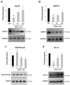
Figure 1
Figure 1
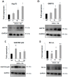
Histamine induces the heat shock response in SOD1-G93A microglia. Representative western blots and quantification of Hsp70 (A), GRP78 (B), SQSTM1/p62 (C) and Bcl-xL (D) in wild-type (WT) and SOD1-G93A microglia treated with histamine (10–100 μM) for 6 h. GAPDH was used for protein normalization. HA = histamine. Data represent mean ± S.E.M. (n = 3 independent experiments). Statistical significance was calculated by ANOVA, as referred to WT cells, # p < 0.05 or to SOD1-G93A untreated cells, * p < 0.05.
Figure 2
Figure 2
Histamine induces the Heat Shock Response in NSC-G93A motor neuron cells. Representative western blots and quantification of Hsp70 (A), GRP78 (B), SQSTM1/p62 (C) and Bcl-xL (D) in differentiated NSC-WT and NSC-G93A cells treated with histamine (10–100 μM) for 48 h. GAPDH was used for protein normalization. HA = histamine. Data represent mean ± S.E.M. (n = 3 independent experiments). Statistical significance was calculated by ANOVA, as referred to NSC-WT cells, # p < 0.05 or to NSC-G93A untreated cells, * p < 0.05; ** p < 0.01. (E) Representative immunofluorescence images of Bcl-xL (green) in NSC-WT and NSC-G93A cells treated with histamine (10–100 μM). Dapi for nuclei staining is shown in blue. Scale bar: 20 μm.
Figure 2
Figure 2
Histamine induces the Heat Shock Response in NSC-G93A motor neuron cells. Representative western blots and quantification of Hsp70 (A), GRP78 (B), SQSTM1/p62 (C) and Bcl-xL (D) in differentiated NSC-WT and NSC-G93A cells treated with histamine (10–100 μM) for 48 h. GAPDH was used for protein normalization. HA = histamine. Data represent mean ± S.E.M. (n = 3 independent experiments). Statistical significance was calculated by ANOVA, as referred to NSC-WT cells, # p < 0.05 or to NSC-G93A untreated cells, * p < 0.05; ** p < 0.01. (E) Representative immunofluorescence images of Bcl-xL (green) in NSC-WT and NSC-G93A cells treated with histamine (10–100 μM). Dapi for nuclei staining is shown in blue. Scale bar: 20 μm.
Figure 3
Figure 3
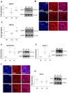
Histidine modulates the expression of Hsp70, GRP78, Beclin-1 and Bcl-xL in the spinal cord of symptomatic SOD1-G93A mice. Representative western blots and quantification of Hsp70 (A), GRP78 (C), SQSTM1/p62 (D), Beclin-1 (E) and Bcl-xL (G) in saline (n = 4) and histidine-treated 100 mg/kg (n = 5) SOD1-G93A mice. GAPDH was used as a loading control. Data represent mean ± S.E.M. Statistical significance was calculated by ANOVA, as referred to WT, # p < 0.05 or to SOD1-G93A saline-treated mice, * p < 0.05. (B) Representative immunofluorescence images of Hsp70 (blue) and SMI32 (red) in saline (n = 4) and 100 mg/kg histidine-treated (n = 5) SOD1-G93A mice. Scale bar: 100 μm. (F) Representative immunofluorescence images of Bcl-xL (blue) and SMI32 (red) in saline (n = 4) and 100 mg/kg histidine-treated (n = 5) SOD1-G93A mice. Scale bar: 100 μm.
Figure 4
Figure 4
Histidine increases Hsp70 and GRP78 and dendritic spine density in the cortex of symptomatic SOD1-G93A mice. Representative western blots and quantification of Hsp70 (A) and GRP78 (B) in saline (n = 4) and 100 mg/kg histidine-treated (n = 5) SOD1-G93A mice. GAPDH was used as a loading control. Representative images of Golgi-stained motor cortex (C), 5× magnification; Scale bar: 50 μm) and of dendritic segments (apical and basal) of Layer V M1 pyramidal neurons from WT, saline (n = 4) and 100 mg/kg histidine-treated (n = 5) SOD1-G93A mice (D), 100× magnification; Scale bar: 5 μm); spine density (n spines/μm) was counted in apical and basal compartment of Layer V pyramidal neurons of M1 (E). Data represent mean ± S.E.M. Statistical significance was calculated by ANOVA, as referred to WT, # p < 0.05 or to SOD1-G93A saline-treated mice, * p < 0.05.
Figure 4
Figure 4
Histidine increases Hsp70 and GRP78 and dendritic spine density in the cortex of symptomatic SOD1-G93A mice. Representative western blots and quantification of Hsp70 (A) and GRP78 (B) in saline (n = 4) and 100 mg/kg histidine-treated (n = 5) SOD1-G93A mice. GAPDH was used as a loading control. Representative images of Golgi-stained motor cortex (C), 5× magnification; Scale bar: 50 μm) and of dendritic segments (apical and basal) of Layer V M1 pyramidal neurons from WT, saline (n = 4) and 100 mg/kg histidine-treated (n = 5) SOD1-G93A mice (D), 100× magnification; Scale bar: 5 μm); spine density (n spines/μm) was counted in apical and basal compartment of Layer V pyramidal neurons of M1 (E). Data represent mean ± S.E.M. Statistical significance was calculated by ANOVA, as referred to WT, # p < 0.05 or to SOD1-G93A saline-treated mice, * p < 0.05.
Figure 5
Figure 5
Synopsis of histamine effects on ALS disease features. In SOD1-G93A motor neurons, histamine increases mitochondrial functionality (ATP content) and survival (SMI32), also protecting axons (SV2/2H3; MBP) [16]. In SOD1-G93A microglia, histamine decreases pro-inflammatory markers (NF-kB, iNOS and NADPH oxidase 2), and increases anti-inflammatory markers (CD163, CD206, IL-6, IL-10 ARG1, P2Y12) together with cell migration [17]. In both motor neurons and microglia histamine activates AKT/ERK1/2 [16,17] in addition to Hsps (GRP78, Hsp70) and Bcl-xL responses, while decreasing SQSTM1/p62 (Figure 1, Figure 2, Figure 3 and Figure 4).

References

    1. Oskarsson B., Gendron T.F., Staff N.P. Amyotrophic Lateral Sclerosis: An Update for 2018. Mayo Clin. Proc. 2018;93:1617–1628. doi: 10.1016/j.mayocp.2018.04.007. - DOI - PubMed
    1. Taylor J.P., Brown R.H., Jr., Cleveland D.W. Decoding ALS: From genes to mechanism. Nature. 2016;539:197–206. doi: 10.1038/nature20413. - DOI - PMC - PubMed
    1. Appel S.H., Zhao W., Beers D.R., Henkel J.S. The microglial-motoneuron dialogue in ALS. Acta Myol. 2011;30:4–8. - PMC - PubMed
    1. Philips T., Robberecht W. Neuroinflammation in amyotrophic lateral sclerosis: Role of glial activation in motor neuron disease. Lancet Neurol. 2011;10:253–263. doi: 10.1016/S1474-4422(11)70015-1. - DOI - PubMed
    1. Zheng Z., Kim J.Y., Ma H., Lee J.E., Yenari M.A. Anti-inflammatory effects of the 70 kDa heat shock protein in experimental stroke. J. Cereb. Blood Flow Metab. 2008;28:53–63. doi: 10.1038/sj.jcbfm.9600502. - DOI - PubMed

MeSH terms

LinkOut - more resources