

Dihydroxy-Substituted Coumarins as Fluorescent Probes for Nanomolar-Level Detection of the 4-Amino-TEMPO Spin Label
- PMID: 31382639
- PMCID: PMC6696051
- DOI: 10.3390/ijms20153802
Dihydroxy-Substituted Coumarins as Fluorescent Probes for Nanomolar-Level Detection of the 4-Amino-TEMPO Spin Label
Abstract
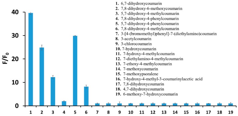
This paper reports on dihydroxycoumarins as fluorescent probes suitable for the detection and determination of the nitroxide radical, namely 4-amino-TEMPO. Since 4-amino-TEMPO is used as a spin label for the detection of various radicals and damage caused by these species, its determination under physiological conditions might help us to understand the mechanism of the oxidative stress. Among different coumarins studied, only dihydroxy-substituted derivatives show high sensitivity, specificity, and selectivity for the nitroxide radical. In this assay, dihydroxy-substituted coumarins under the action of 4-amino-TEMPO show a very fast and significant increase in fluorescence intensity and lifetime. Among them 6,7-dihydroxycoumarin (esculetin) exhibits the strongest fluorescence enhancement (up to 40 times), with an estimated limit of detection equal to 16.7 nM-a significantly lower value when compared with UV-Vis or electron paramagnetic resonance (EPR) spectroscopy. The method is characterized by an easy procedure of sample preparation and very short time of analysis. The mechanism of the interaction between 6,7-dihydroxycoumarin and 4-amino-TEMPO has been examined with the use of a series of complementary techniques, such as steady-state and time-resolved fluorescence spectroscopy, UV-Vis spectroscopy, electron paramagnetic resonance spectroscopy, potentiometric titration, and high-performance liquid chromatography. It has been proven that the only route of the reaction in the system studied is a proton transfer from the molecule of esculetin to the amino group of the nitroxide. Biological studies performed on prostate cancer cells, breast cancer cells, and normal skin fibroblasts revealed significant anticancer properties of 6,7-dihydroxycoumarin, which caused a considerable decrease in the viability and number of cancer cells, and affected their morphology, contrary to normal fibroblasts. Furthermore, the experiment performed on prostate cancer cells showed that fluorescence emission of esculetin is closely related to intracellular pH-the higher pH, the higher observed fluorescence intensity (in accordance with a chemical experiment). On the other hand, the studies performed in different pH levels revealed that when pH of the solution increases, the observed fluorescence intensity enhancement under the action of 4-amino-TEMPO decreases (better sensing properties of esculetin towards the nitroxide in lower pH).
Keywords: dihydroxycoumarins; fluorescence; nitroxide radicals; optical chemical sensors.
Conflict of interest statement
The authors declare no conflict of interest.
Figures


References
-
- Żamojć K., Zdrowowicz M., Rudnicki-Velasquez P.B., Krzymiński K., Zaborowski B., Niedziałkowski P., Jacewicz D., Chmurzyński L. The development of 1,3-diphenylisobenzofuran as a highly selective probe for the detection and quantitative determination of hydrogen peroxide. Free Radic. Res. 2017;51:38–46. doi: 10.1080/10715762.2016.1262541. - DOI - PubMed
-
- Żamojć K., Jacewicz D., Zdrowowicz M., Chmurzyński L. Kinetics of the reaction between 1,3-diphenylisobenzofuran and nitrogen dioxide studied by steady-state fluorescence. Res. Chem. Intermed. 2013;39:3023–3031. doi: 10.1007/s11164-012-0814-4. - DOI
MeSH terms
Substances
Grants and funding
LinkOut - more resources
Full Text Sources
