. 2019 Aug 5;20(1):29.
doi: 10.1186/s12860-019-0213-4.

CD40/anti-CD40 antibody complexes which illustrate agonist and antagonist structural switches

Affiliations

CD40/anti-CD40 antibody complexes which illustrate agonist and antagonist structural switches

Maria A Argiriadi et al. BMC Mol Cell Biol. .

Abstract

Background: CD40 is a 48 kDa type I transmembrane protein that is constitutively expressed on hematopoietic cells such as dendritic cells, macrophages, and B cells. Engagement of CD40 by CD40L expressed on T cells results in the production of proinflammatory cytokines, induces T helper cell function, and promotes macrophage activation. The involvement of CD40 in chronic immune activation has resulted in CD40 being proposed as a therapeutic target for a range of chronic inflammatory diseases. CD40 antagonists are currently being explored for the treatment of autoimmune diseases and several anti-CD40 agonist mAbs have entered clinical development for oncological indications.

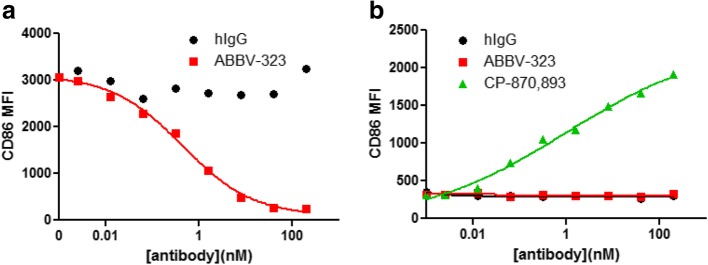
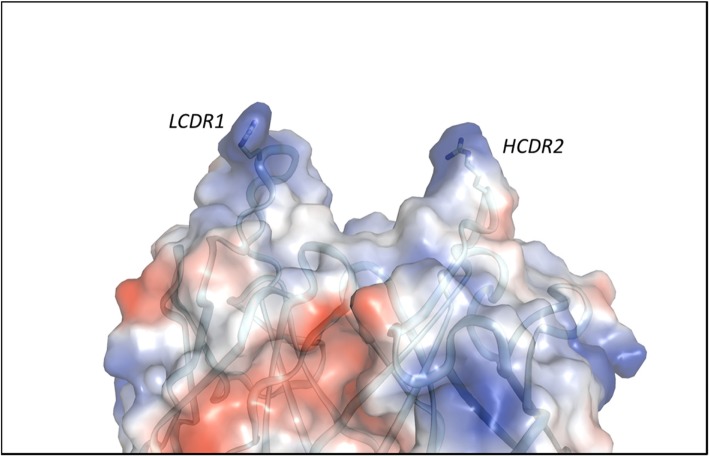
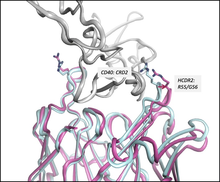
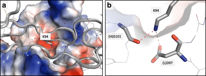
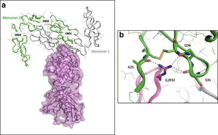
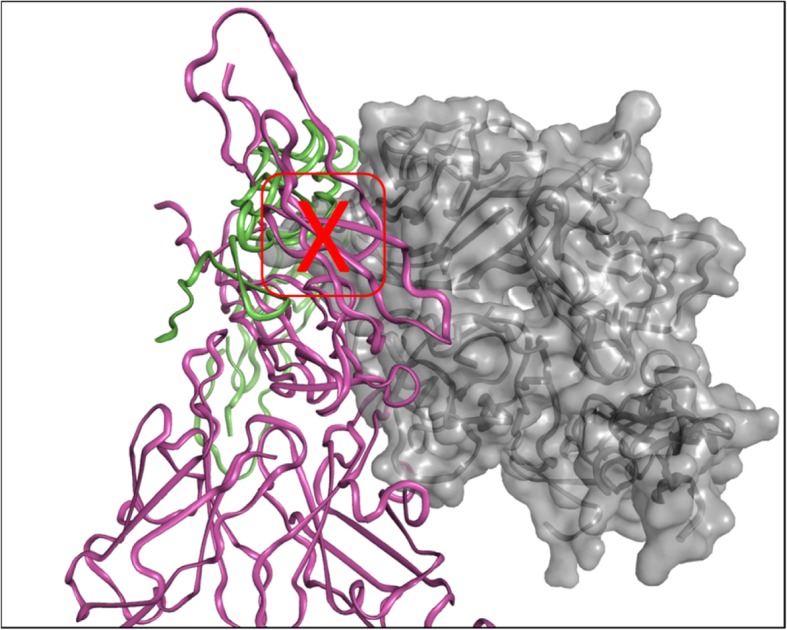
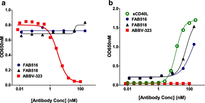
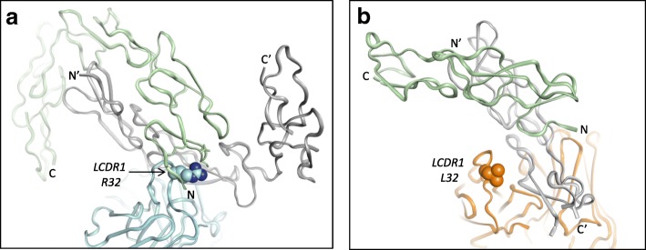
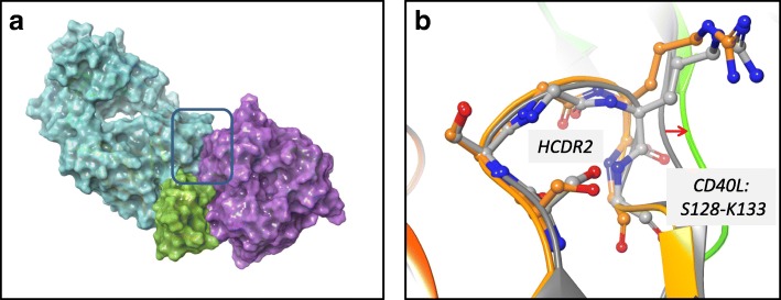
Results: To better understand the mode of action of anti-CD40 mAbs, we have determined the x-ray crystal structures of the ABBV-323 (anti-CD40 antagonist, ravagalimab) Fab alone, ABBV-323 Fab complexed to human CD40 and FAB516 (anti-CD40 agonist) complexed to human CD40. These three crystals structures 1) identify the conformational CD40 epitope for ABBV-323 recognition 2) illustrate conformational changes which occur in the CDRs of ABBV-323 Fab upon CD40 binding and 3) develop a structural hypothesis for an agonist/antagonist switch in the LCDR1 of this proprietary class of CD40 antibodies.

Conclusions: The structure of ABBV-323 Fab demonstrates a unique method for antagonism by stabilizing the proposed functional antiparallel dimer for CD40 receptor via novel contacts to LCDR1, namely residue position R32 which is further supported by a closely related agonist antibody FAB516 which shows only monomeric recognition and no contacts with LCDR1 due to a mutation to L32 on LCDR1. These data provide a structural basis for the full antagonist activity of ABBV-323.

Keywords: Agonist; Antagonist; Antibody; CD40; Crystal structure.


Conflict of interest statement

The authors declare that they have no competing interests.

Figures

Fig. 1
Fig. 1
a and b: (a) ABBV-323 strongly inhibits CD40 signaling in B cells (inhibition of CD86 expression) without inducing agonist activity (stimulation of CD86 expression). To measure antagonist activity, CD40L+ jurkat cells were used to stimulate primary human B cells +/− ABBV-323. (b) To measure agonist activity, B cells were incubated with anti-CD40 antibodies
Fig. 2
Fig. 2
Electrostatic potential surface calculated in Pymol for Fab ABBV-323 crystal structure. A cleft is formed between HCDR2 and LCDR1. Red patches refer to negative charged regions and blue patches refer to positive charged regions
Fig. 3
Fig. 3
Complex structure of ABBV-323 Fab (magenta) and human CD40 (shown in grey). The Fab alone structure is superimposed (in cyan) to show how HCDR2 opens to accommodate CD40
Fig. 4
Fig. 4
a and b: (a) K94 inserts into a negatively charged channel to make interactions with acidic pocket. (b) Interactions between K94 and (H)G101/(L)D97 are illustrated
Fig. 5
Fig. 5
a and b: (a) Two crystallographic CD40 monomers associate to form a tight antiparallel dimer (grey, green) which bind to ABBV-323 Fab (shown in magenta). (b) LCDR1 R32 inserts in the second crystallographic monomer shown in green. R32 makes interactions with backbone carbonyls to A25/S35 and sidechain Q36
Fig. 6
Fig. 6
Overlay of ABBV-323/CD40 complex (magenta = Fab/CD40 monomer 1, green = CD40 crystallographic monomer 2) with crystal structure of CD40L/CD40 complex (PDB code: 3QD6 in grey surface) [4]. Red outlined box shows the region of significant steric clash between CD40 dimer and CD40L
Fig. 7
Fig. 7
a and b: (a) ABBV-323 was incubated with HEK-293 CD40L cells in the presence of CD40L expressing D1.1 cells. The ability of ABBV-323 to inhibit the interaction between CD40 and its receptor CD40L is monitored by the production of SEAP compared to an IgG control. (b) Agonist activity is monitored as above with the following exception: CD40L-expressing D1.1 cells were replaced with assay media. CD40L was titrated alongside ABBV-323 as a positive control (Concentration range = 250 ng/ml – 0.026 ng/ml)
Fig.8
Fig.8
a and b (a) ABBV-323 Fab (blue) bound to crystallographic CD40 dimer (second monomer shown in green ribbon) which shows interaction with LCDR1 R32. (b) FAB516 Fab (orange) bound to crystallographic CD40 dimer (second monomer shown in green ribbon) which shows no interaction with LCDR1 L32. N and C termini are also labeled to demonstrate the antiparallel dimers for both ABBV-323 and FAB516
Fig. 9
Fig. 9
a and b: (a) Model of potential ternary complex of agonist FAB516 Fab (cyan), human CD40 (green), human CD40L (purple). Box outlines region where minor structural rearrangements may occur to accommodate agonist complex. (b) After ternary complex minimization in MAESTRO, HCDR2 (S52-G56) and CD40L (S128-K132) residues shift to accommodate the interface between CD40L, FAB516 and CD40

References

    1. Bishop GA, Moore CR, Xie P, Stunz LL, Kraus ZJ. TRAF proteins in CD40 signaling. Adv Exp Med Biol. 2007;597:131–151. doi: 10.1007/978-0-387-70630-6_11. - DOI - PubMed
    1. Hostager BS, Haxhinasto SA, Rowland SL, Bishop GA. Tumor necrosis factor receptor-associated factor 2 (TRAF2)-deficient B lymphocytes reveal novel roles for TRAF2 in CD40 signaling. J Biol Chem. 2007;278:45382–45390. doi: 10.1074/jbc.M306708200. - DOI - PubMed
    1. Law CL, Grewal IS. Therapeutic interventions targeting CD40L (CD154) and CD40: the opportunities and challenges. Adv Exp Med Biol. 2009;647:8–36. doi: 10.1007/978-0-387-89520-8_2. - DOI - PubMed
    1. An HJ, Kim YJ, Song DH, Park BS, Kim HM, Lee JD, Paik SG, Lee JO, Lee H. Crystallographic and mutational analysis of the CD40-CD154 complex and its implications for receptor activation. J Biol Chem. 2011;286:11226–11235. doi: 10.1074/jbc.M110.208215. - DOI - PMC - PubMed
    1. Koyama I, Kawai T, Andrews D, Boskovic S, Nadazdin O, Wee SL, Sogawa H, Wu DL, Smith RN, Colvin RB, Sachs DH, Cosimi AB. Thrombophilia associated with anti-CD154 monoclonal antibody treatment and its prophylaxis in nonhuman primates. Transplantation. 2004;77:460–462. doi: 10.1097/01.TP.0000110291.29370.C0. - DOI - PubMed