Case Reports
. 2019 Aug 5;17(1):134.
doi: 10.1186/s12957-019-1681-x.

Needle tract seeding following endoscopic ultrasound-guided fine-needle aspiration for pancreatic cancer: a report of two cases

Affiliations
Case Reports

Needle tract seeding following endoscopic ultrasound-guided fine-needle aspiration for pancreatic cancer: a report of two cases

Toshiki Matsui et al. World J Surg Oncol. .

Abstract

Background: Endoscopic ultrasound-guided fine-needle aspiration (EUS-FNA) is a useful tool in pancreatic cancer diagnosis. However, the procedure itself may cause peritoneal dissemination and needle tract seeding at the puncture site. We herein report two cases of gastric wall metastasis due to needle tract seeding after EUS-FNA.

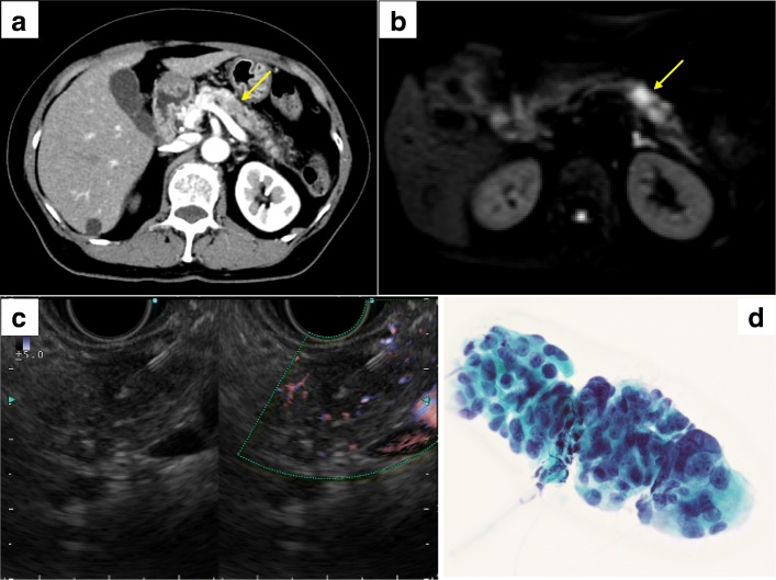
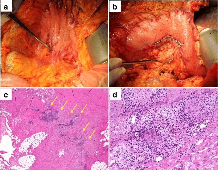
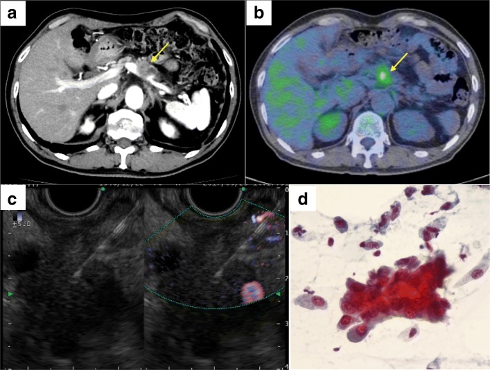
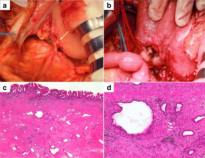
Case presentation: Case 1: A 68-year-old woman was admitted to our hospital for persistent cough. Computed tomography (CT) scan revealed inflammatory changes in the left lung field, and incidentally, a 15-mm hypovascular mass was detected in the pancreatic body. She underwent EUS-FNA and was diagnosed as pancreatic adenocarcinoma. She underwent distal pancreatectomy with splenectomy; however, a small hard mass was observed in the posterior gastric wall during surgery. We performed partial gastrectomy, and the resected specimen was diagnosed as a needle tract seeding following EUS-FNA. She then underwent adjuvant chemotherapy with TS-1, but the pancreatic cancer showed recurrence 6 months after surgery. She died due to peritoneal dissemination 18 months after surgery. Case 2: A 70-year-old man was incidentally detected with a pancreatic body mass on a CT scan as part of his follow-up for recurrence of basal cell carcinoma. He underwent EUS-FNA and was diagnosed as pancreatic adenocarcinoma. He had nodules in both lungs, and it was difficult to differentiate them from lung metastasis of pancreatic cancer. Therefore, he underwent neoadjuvant chemoradiotherapy, and thereafter, the lung nodules showed no changes; hence, he underwent distal pancreatectomy with splenectomy. During surgery, we observed a hard mass in the posterior gastric wall. We performed partial gastrectomy, and the resected specimen was diagnosed as needle tract seeding due to EUS-FNA. He underwent chemotherapy with TS-1, and he is still alive 18 months after surgery at the time of writing.

Conclusion: For resectable pancreatic body or tail tumors, EUS-FNA should be carefully performed to prevent needle tract seeding and intraoperative as well as postoperative assessment for gastric wall metastasis is mandatory.

Keywords: Endoscopic ultrasound-guided fine-needle aspiration; Gastric wall metastasis; Needle tract seeding; Pancreatic cancer; Surgical resection.


Conflict of interest statement

The authors declare that they have no competing interests.

Figures

Fig. 1
Fig. 1
a Dynamic-enhanced computed tomography (portal phase) for case 1. A 15-mm hypovascular tumor was detected in the pancreatic body (arrow). b Diffusion-weighted magnetic resonance imaging. A hyperintense area can be observed in the pancreatic body tumor (arrow). c Endoscopic ultrasound-guided fine-needle aspiration (EUS-FNA). EUS-FNA was performed for the pancreatic tumor (4 punctures using 22 G, 19 G, 20 G, and 20 G needles) via the trans-gastric approach, without any complications. d Pathological findings of EUS-FNA. An adenocarcinoma can be observed (Papanicolaou staining)
Fig. 2
Fig. 2
a Intraoperative findings for case 1. A small hard mass was detected in the posterior gastric wall, as indicated by the forceps. b Partial resection of the posterior gastric wall was performed. c Pathological findings. The specimen from the partially resected stomach showed that an adenocarcinoma was distributed linearly in the gastric muscle layer (arrow) (hematoxylin and eosin staining, loupe image). d Pathological findings. The findings of the gastric tumor were similar to those of the primary pancreatic cancer, indicating that gastric tumor was needle tract seeding from pancreatic cancer (hematoxylin and eosin staining)
Fig. 3
Fig. 3
a Dynamic-enhanced CT (portal phase) for case 2. A 15-mm hypovascular tumor in the pancreatic body (arrow). b Positron emission tomography-CT (PET-CT) findings. Abnormal accumulation of fluorine-18-deoxyglucose (standardized uptake value of 3.74) can be observed in the pancreatic body (arrow). c EUS-FNA findings. EUS-FNA was performed for the pancreatic tumor (1 puncture using 22 G, needle) via the trans-gastric approach, without any complications. d Pathological findings. EUS-FNA revealed an adenocarcinoma (Papanicolaou staining)
Fig. 4
Fig. 4
a Intraoperative findings for case 2. A small hard mass was detected in the posterior gastric wall (arrow). b Partial resection of the posterior gastric wall was performed. c Pathological findings. Many abnormal luminal structures (adenocarcinoma) were confirmed in the resected gastric muscle layer (hematoxylin and eosin staining, loupe image). d Pathological findings. The findings of gastric tumor were similar to those of the primary pancreatic cancer, indicating that gastric tumor was a recurrence due to needle tract seeding from pancreatic cancer (hematoxylin and eosin staining)

Similar articles

Cited by

References

    1. Hewitt MJ, McPhail MJW, Possamai L, Dhar A, Vlavianos P, Monahan KJ. EUS-guided FNA for diagnosis of solid pancreatic neoplasms: a meta-analysis. Gastrointest Endosc. 2012;75:319–331. doi: 10.1016/j.gie.2011.08.049. - DOI - PubMed
    1. Wang KX, Ben QW, Jin ZD, Du YQ, Zou DW, Liao Z, et al. Assessment of morbidity and mortality associated with EUS-guided FNA: a systematic review. Gastrointest Endosc. 2011;73:283–290. doi: 10.1016/j.gie.2010.10.045. - DOI - PubMed
    1. Micames C, Jowell PS, White R, Paulson E, Nelson R, Morse M, et al. Lower frequency of peritoneal carcinomatosis in patients with pancreatic cancer diagnosed by EUS-guided FNA vs. percutaneous FNA. Gastrointest Endosc. 2003;58:690–695. doi: 10.1016/S0016-5107(03)02009-1. - DOI - PubMed
    1. Yamauchi J, Kobayashi S, Miyazaki K, Ajiki T, Tsuchihara K, Ishiyama S. A case of curative resection of needle tract seeding after EUS-guided fine needle aspiration for pancreatic body cancer. J Jpn Surg Assoc. 2016;77(12):2994–2999. doi: 10.3919/jjsa.77.2994. - DOI
    1. Hirooka Y, Goto H, Itoh A, Hashimoto S, Niwa K, Ishikawa H, et al. Case of intraductal papillary mucinous tumor in which endosonograpy-guided fine-needle aspiration biopsy caused dissemination. J Gastroenterology Hepatol. 2003;18:1323–1324. doi: 10.1046/j.1440-1746.2003.03040.x. - DOI - PubMed

Publication types