. 2019 Aug 5;10(1):236.
doi: 10.1186/s13287-019-1354-2.

Modulation of Wnt/BMP pathways during corneal differentiation of hPSC maintains ABCG2-positive LSC population that demonstrates increased regenerative potential

Affiliations

Modulation of Wnt/BMP pathways during corneal differentiation of hPSC maintains ABCG2-positive LSC population that demonstrates increased regenerative potential

Meri Vattulainen et al. Stem Cell Res Ther. .

Abstract

Background: The differentiation of corneal limbal stem cells (LSCs) from human pluripotent stem cells (hPSCs) has great power as a novel treatment for ocular surface reconstruction and for modeling corneal epithelial renewal. However, the lack of profound understanding of the true LSC population identity and the regulation of LSC homeostasis is hindering the full therapeutic potential of hPSC-derived LSCs as well as primary LSCs.

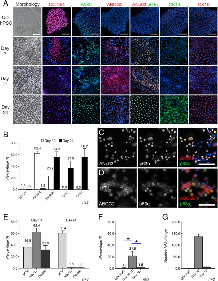
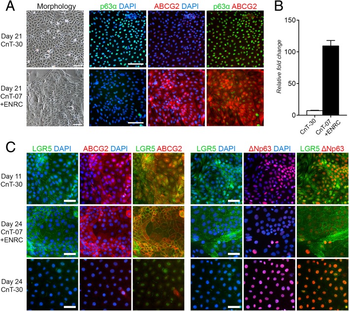
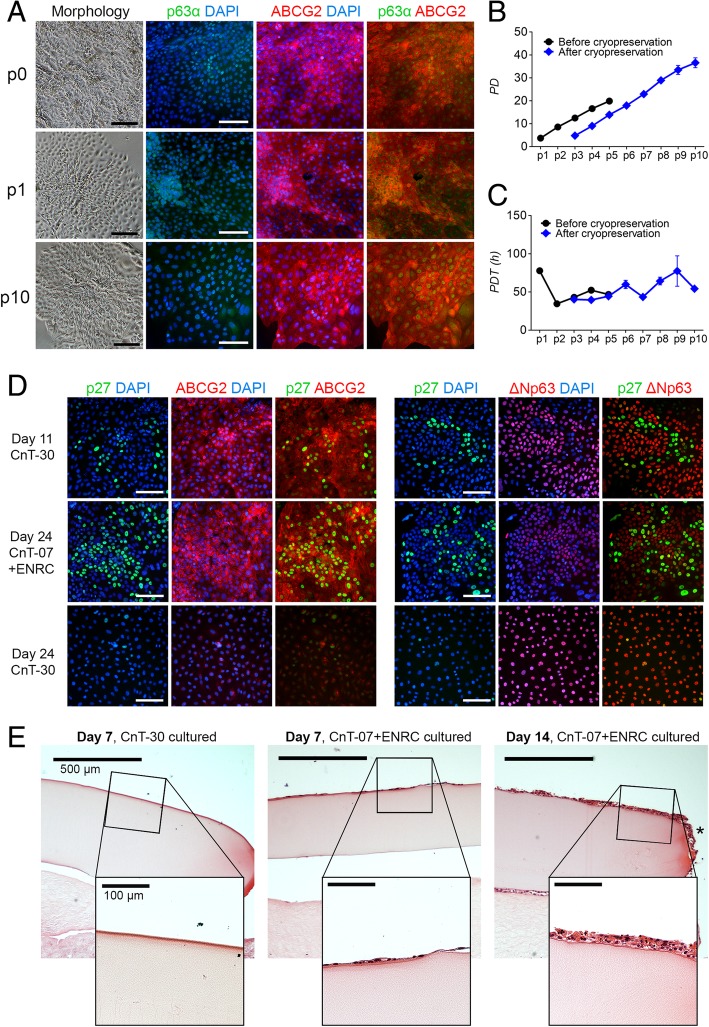
Methods: The differentiation trajectory of two distinct hPSC lines towards LSCs was characterized extensively using immunofluorescence labeling against pluripotency, putative LSC, and mature corneal epithelium markers. Cell counting, flow cytometry, and qRT-PCR were used to quantify the differences between distinct populations observed at day 11 and day 24 time points. Initial differentiation conditions were thereafter modified to support the maintenance and expansion of the earlier population expressing ABCG2. Immunofluorescence, qRT-PCR, population doubling analyses, and transplantation into an ex vivo porcine cornea model were used to analyze the phenotype and functionality of the cell populations cultured in different conditions.

Results: The detailed characterization of the hPSC differentiation towards LSCs revealed only transient expression of a cell population marked by the universal stemness marker and proposed LSC marker ABCG2. Within the ABCG2-positive population, we further identified two distinct subpopulations of quiescent ∆Np63α-negative and proliferative ∆Np63α-positive cells, the latter of which also expressed the acknowledged intestinal stem cell marker and suggested LSC marker LGR5. These populations that appeared early during the differentiation process had stem cell phenotypes distinct from the later arising ABCG2-negative, ∆Np63α-positive third cell population. Importantly, novel culture conditions modulating the Wnt and BMP signaling pathways allowed efficient maintenance and expansion of the ABCG2-positive populations. In comparison to ∆Np63α-positive hPSC-LSCs cultured in the initial culture conditions, ABCG2-positive hPSC-LSCs in the novel maintenance condition contained quiescent stem cells marked by p27, demonstrated notably higher population doubling capabilities and clonal growth in an in vitro colony-forming assay, and increased regenerative potential in the ex vivo transplantation model.

Conclusions: The distinct cell populations identified during the hPSC-LSC differentiation and ABCG2-positive LSC maintenance may represent functionally different limbal stem/progenitor cells with implications for regenerative efficacy.

Keywords: ABCG2; Human pluripotent stem cells; Limbal stem cell deficiency; Limbal stem cells; Stem cell differentiation; Stem cell hierarchy; Stem cell maintenance; Wnt signaling.


Conflict of interest statement

Tampere University Finland has a patent pending regarding the novel hPSC-LSC maintenance method fully reported herein. Based on the Act on the Right in Inventions made at Higher Education Institutions in Finland, all authors employed by Tampere University have given all rights to the University and thus the authors declare that they have no competing interests.

Figures

Fig. 1
Fig. 1
Flow chart of the experimental design and progression. a Standard CnT-30-based hPSC-LSC differentiation protocol and characterization of the hPSC-LSC differentiation process. b Novel CnT-07+ENRC-based hPSC-LSC maintenance protocol, characterization, and comparison of distinct cell populations identified during the study. PSC pluripotent stem cell, UD-hPSC undifferentiated human PSC, LSC limbal stem cell, IF immunofluorescence, qRT-PCR quantitative real-time PCR, LN-521 laminin-521, Col IV collagen type IV, E8 Flex, E8 Flex pluripotent stem cell culture medium, CnT-30 CnT-30 corneal differentiation medium, CnT-07 CnT-07 epithelial proliferation medium, ENRC epidermal growth factor, Noggin, R-Spondin-1, CHIR99021
Fig. 2
Fig. 2
Characterization of putative LSC marker expression during hPSC-LSC differentiation. a Representative morphology and protein expression of the cultures at selected time points. Scale bars, 100 μm for all images in the same column. Cell nuclei counterstained with DAPI (blue). b Marker expression differences in the d10 and d24 populations. Five images per sample and a minimum of 1400 cells per time point were analyzed for each marker from cytospin samples. c Representative IF image of ∆Np63 and p63α double-staining in a d24 cytospin sample. Scale bar, 100 μm for both c and d. d Representative IF image of ABCG2 and p63α double-staining in a d10 cytospin sample. e p63α and ABCG2 expression in d10 and d24 hPSC-LSCs. Five images per sample and a minimum of 3 000 cells per time point were analyzed from cytospin samples. P > 0.05. f The level of ABCG2 protein expression in UD-hPSCs and in d10 and d24–26 hPSC-LSCs, analyzed with flow cytometry. g The ABCG2 mRNA expression levels in UD-hPSCs and in d10 and d24 hPSC-LSCs analyzed with qRT-PCR. All representative data are presented with the hESC line Regea08/017. All quantitative data are presented as the mean + SD, and n marks the individual cell differentiation batches serving as biological replicates. Statistical analyses were carried out using the Mann-Whitney U test. *P ≤ 0.05
Fig. 3
Fig. 3
CD200 and ABCG2 expression patterns during differentiation of hPSC towards LSCs, analyzed with flow cytometry. Scatter plots and adjunct histograms as well as tables showing the distribution of cells in the CD200+/ABCG2 (Q1), CD200+/ABCG2+ (Q2), CD200/ABCG2+ (Q3), and CD200/ABCG2 (Q4) subpopulations of UD-hPSCs (a) as well as at d11 (b) and d29 (c) during hPSC-LSC differentiation. For each sample, 10,000 initially gated events were analyzed, and the experiment was carried out once for the representative line Regea08/017
Fig. 4
Fig. 4
Effect of culture conditions on hPSC-LSC morphology and p63α, ABCG2, and LGR5 expression. a Representative cell morphology and p63α/ABCG2 protein expression under different culture conditions, as demonstrated by IF. Scale bars, 100 μm. b ABCG2 mRNA expression under different conditions, analyzed with qRT-PCR in d21 cells. Data are presented as the mean + SD, n = 3 technical replicates from one sample, P > 0.05. c Characterization of LGR5 protein expression in relation to ABCG2 and ∆Np63 at d11, as well as after continued culture in CnT-07+ENRC or CnT-30 at d24. Cell nuclei counterstained with DAPI (blue). Scale bars, 50 μm. Data shown for the representative hESC line Regea08/017
Fig. 5
Fig. 5
Marker expression and functional proliferative and regenerative properties of the ABCG2-positive hPSC-LSCs cultured in the novel maintenance condition. a Morphology and p63α/ABCG2 expression pattern of hPSC-LSCs cultured in CnT-07+ENRC up to passages 0, 1, and 10. Cell nuclei counterstained with DAPI (blue). Scale bars, black = 200 μm, white = 100 μm. b Population doublings and c population doubling times of freshly differentiated hPSC-LSCs up to passage 5 and cryopreserved hPSC-LSCs up to passage 10. d Characterization of expression of the quiescence marker p27 in relation to ABCG2 and ∆Np63 at d11, as well as after continued culture in CnT-07+ENRC or CnT-30 at d24. Scale bars, 100 μm. e Hematoxylin and eosin-stained sections of CnT-30-cultured Regea08/017 and CnT-07+ENRC-cultured Regea11/013 hPSC-LSCs after 1-week and 2-week time points in the ex vivo porcine cornea model, respectively. Asterisk points out migrating cells. Scale bars, 500 μm and 100 μm, for all images in the panel. Data are presented for the hESC lines Regea11/013 (ac, e) and Regea08/017 (d, e)
Fig. 6
Fig. 6
Proposed differentiation hierarchy of the LSC populations in the human cornea. a Schematic illustration of the human limbus, proposing that populations of both quiescent (purple) and actively cycling (green) LSCs coexist in the limbal crypts, from where their TAC (blue) progeny migrate towards the central cornea, going through several cell divisions before terminal differentiation into mature (white) CECs. b Representation of the interplay between separate limbal cell populations, identified by distinct marker expression profiles. LSC limbal stem cell, TAC transiently amplifying cell, CEC corneal epithelial cell

Similar articles

Cited by

References

    1. Dua HS, Shanmuganathan VA, Powell-Richards AO, et al. Limbal epithelial crypts: a novel anatomical structure and a putative limbal stem cell niche. Br J Ophthalmol. 2005;89:529–532. doi: 10.1136/bjo.2004.049742. - DOI - PMC - PubMed
    1. Rama P, Ferrari G, Pellegrini G. Cultivated limbal epithelial transplantation. Curr Opin Ophthalmol. 2017;28:387–389. doi: 10.1097/ICU.0000000000000382. - DOI - PubMed
    1. Chakrabarty K, Shetty R, Ghosh A. Corneal cell therapy: with iPSCs, it is no more a far-sight. Stem Cell Res Ther. 2018;9:1–15. doi: 10.1186/s13287-018-1036-5. - DOI - PMC - PubMed
    1. Haagdorens M, Van Acker SI, Van Gerwen V, et al. Limbal stem cell deficiency: current treatment options and emerging therapies. Stem Cells Int. 2016;2016:1–22. doi: 10.1155/2016/9798374. - DOI - PMC - PubMed
    1. Schlötzer-Schrehardt U, Kruse FE. Identification and characterization of limbal stem cells. Exp Eye Res. 2005;81:247–264. doi: 10.1016/j.exer.2005.02.016. - DOI - PubMed

Publication types

MeSH terms

Substances