. 2020 Jan:106:102306.
doi: 10.1016/j.jaut.2019.102306. Epub 2019 Aug 2.

Restricted myeloperoxidase epitopes drive the adaptive immune response in MPO-ANCA vasculitis

Affiliations

Restricted myeloperoxidase epitopes drive the adaptive immune response in MPO-ANCA vasculitis

Meghan E Free et al. J Autoimmun. 2020 Jan.

Abstract

Background: Treatment of autoimmune diseases has relied on broad immunosuppression. Knowledge of specific interactions between human leukocyte antigen (HLA), the autoantigen, and effector immune cells, provides the foundation for antigen-specific therapies. These studies investigated the role of HLA, specific myeloperoxidase (MPO) epitopes, CD4+ T cells, and ANCA specificity in shaping the immune response in patients with anti-neutrophil cytoplasmic autoantibody (ANCA) vasculitis.

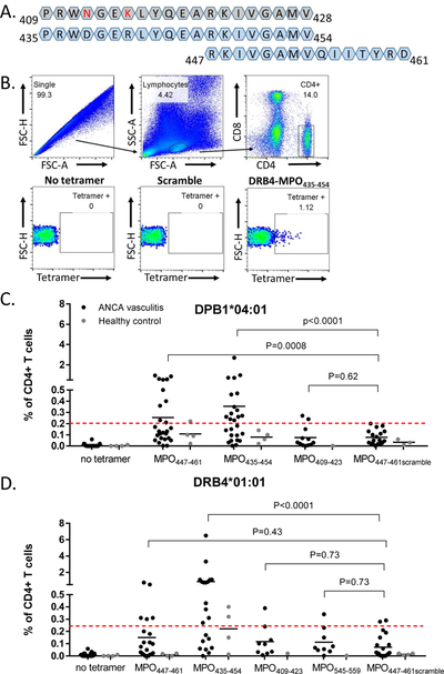
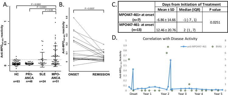
Methods: HLA sequence-based typing identified enriched alleles in our patient population (HLA-DPB1*04:01 and HLA-DRB4*01:01), while in silico and in vitro binding studies confirmed binding between HLA and specific MPO epitopes. Class II tetramers with MPO peptides were utilized to detect autoreactive CD4+ T cells. TCR sequencing was performed to determine the clonality of T cell populations. Longitudinal peptide ELISAs assessed the temporal nature of anti-MPO447-461 antibodies. Solvent accessibility combined with chemical modification determined the buried regions of MPO.

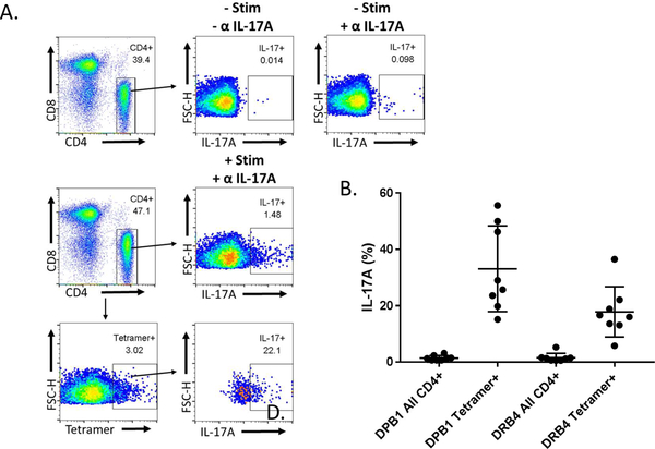
Results: We identified a restricted region of MPO that was recognized by both CD4+ T cells and ANCA. The autoreactive T cell population contained CD4+CD25intermediateCD45RO+ memory T cells and secreted IL-17A. T cell receptor (TCR) sequencing demonstrated that autoreactive CD4+ T cells had significantly less TCR diversity when compared to naïve and memory T cells, indicating clonal expansion. The anti-MPO447-461 autoantibody response was detectable at onset of disease in some patients and correlated with disease activity in others. This region of MPO that is targeted by both T cells and antibodies is not accessible to solvent or chemical modification, indicating these epitopes are buried.

Conclusions: These observations reveal interactions between restricted MPO epitopes and the adaptive immune system within ANCA vasculitis that may inform new antigen-specific therapies in autoimmune disease while providing insight into immunopathogenesis.

Keywords: ANCA specificity; ANCA vasculitis; Autoreactive T cells; Epitope specificity; Immunodominant epitopes.


Conflict of interest statement

The authors have declared that no conflict of interest exists.

Figures

Figure 1.
Figure 1.
Patients demonstrate HLA and epitope specific tetramer recognition by CD4+ T cells. (A) Amino acid sequence alignment of mouse MPO409–428 (grey), human MPO435–454, and human MPO447–461. Amino acid differences between mouse and human epitopes in red. (B) Gating strategy for tetramer+CD4+ T cells with no tetramer control, MPO447–461 scramble control, and HLA-DRB4 MPO435–454. Tetramer positivity for each individual was based on the corresponding no-tetramer control. (C-D) CD4+ T cell recognition of tetramers in MPO-ANCA patients (n=27; black dots) and HLA-matched healthy controls (n=6; gray dots). Some patients and healthy controls had both HLAs and were used in both studies while some samples were only used for one HLA. Dashed red line indicates threshold of positivity determined by the average plus two standard deviations of patient and healthy control MPO447–461scramble tetramer positivity. (C) HLA-DPB1*04:01 tetramer carrying MPO peptides (D) HLA-DRB4*01:01 tetramers carrying MPO peptides. P values were calculated by paired signed rank test.
Figure 2.
Figure 2.
Patient CD4+ T cells that bind tetramers are enriched for CD25intermediate cells expressing CD45RO, indicating memory cells memory marker (CD45RO+). (A) Gating strategy for CD25intermediate cells using a representative patient. Consistently half of tetramer positive cells were CD25intermediate T cells (n=14). (B) Representative plot demonstrating that the expression of CD45RO+ was slightly higher in tetramer positive cells than overall CD4+ T cells (n=6).
Figure 3.
Figure 3.
Tetramer positive T cells are enriched for cells capable of IL-17A secretion. (A) Representative plot shows gating scheme for determining percentage of IL-17A secreting cells for all CD4+ and tetramer positive cells in patients compared to no stimulation and no antibody (α IL-17A) controls. (B) Compared to all CD4+ T cells, a higher proportion of tetramer positive CD4+ T cells secrete IL-17A in patients (n=8)
Figure 4.
Figure 4.
Decreased diversity of MPO435–454 specific T cells (Tet+) relative to tetramer negative CD25, CD25intermediate, and regulatory T cells. Matched bootstrapped diversity including species richness, Shannon entropy, and d50 diversity indices derived from T cell receptor beta (TRB) profiling are shown (n=5 patients, n=4 for tetramer positive and Treg samples) corrected for false discovery rate (FDR). * p<0.05
Figure 5.
Figure 5.
Anti-MPO447–461 autoantibody is detectable in 53% of patients tested by ELISA (n=27/51), occasionally at disease onset. (A) Measured serum or plasma anti-MPO447–461 ELISA reactivity from healthy controls (HC), patients with PR3-ANCA vasculitis, patients with systemic lupus erythematous (SLE), and patients with MPO-ANCA vasculitis, normalized to a positive control. Threshold of positivity was determined by the average of healthy control samples plus two standard deviations and is indicated by the grey dashed line. P-values calculated using Wilcoxon two sample test with a Bonferroni adjusted critical value of 0.0083. (B) Reactivity of 20 samples collected at onset paired with a remission sample from the same patient, normalized to a positive control. P-value calculated using matched-pairs sign rank test (C) Analysis of onset samples revealed that anti-MPO447–461 negative samples had a higher prevalence of treatment prior to collection. P-values calculated using Wilcoxon two sample test. (D) Representative patient where temporal analysis of patient anti-MPO447–461 reactivity correlates with disease activity (BVAS score).
Figure 6.
Figure 6.
Anti-MPO447–461 antibody binding is dependent on secondary structure. (A) Alanine scanning experiments were performed in triplicate with eight previously positive patients by ELISA. Each letter represents the amino acid switched for alanine in sequential peptides. The positive control was the same highly positive serum included on every plate and the same healthy control was included on every plate. Scramble is the MPO447–461 scrambled control and a secondary antibody only control was also used. (B) Circular dichroism experiments to evaluate the secondary structure of peptides with alanine substitutions. The spectra were the average of four scans obtained by collecting data at 0.2 nm intervals from 260 to 190 nm, with a response time of 2 seconds and a bandwidth of 1 nm. (C) Cartoon model of MPO447–461 highlighted in blue demonstrate alpha-helical structure of the region. All models were generated using Pymol.
Figure 7.
Figure 7.
Solvent accessibility and chemical modification determine the buried nature of MPO447–461. (A) Accessibility to solvent of each amino acid in MPO447–459 determined by predictive algorithms. Residues are considered to be solvent exposed if ratio value > 50%, and buried if the ratio value < 20%. (B) Isotope labeling with succinic anhydride to determine solvent accessibility of MPO epitopes. The top panel examines the percentage of succinylation versus the accessibility of ε-NH2 Lysine and N-terminal α-NH2. MPO447–461 ((R)KIVGAMVQIITYR(D)), represented by the circled data point, had almost undetectable levels of succinylation and solvent accessibility.
Figure 8.
Figure 8.
Human MPO crystal structure reveals overlap between previously reported MPO epitopes. (PDBID:5FIW) Highlighted regions are as follows; dark blue - MPO435–454 (Ooi et al.2012), purple- MPO447–459 (Roth et al. 2013), light blue - MPO457–465 (Chang et al. 2017), gold - overlap between highlighted regions. The active site heme of myeloperoxidase is colored red.

Similar articles

Cited by

References

    1. Lyons PA, Rayner TF, Trivedi S, Holle JU, Watts RA, Jayne DR, Baslund B, Brenchley P, Bruchfeld A, Chaudhry AN, et al. 2012. Genetically distinct subsets within ANCA-associated vasculitis. N Engl J Med 367:214–223. - PMC - PubMed
    1. Xie G, Roshandel D, Sherva R, Monach PA, Lu EY, Kung T, Carrington K, Zhang SS, Pulit SL, Ripke S, et al. 2013. Association of granulomatosis with polyangiitis (Wegener’s) with HLA-DPB1*04 and SEMA6A gene variants: evidence from genome-wide analysis. Arthritis Rheum 65:2457–2468. - PMC - PubMed
    1. Merkel PA, Xie G, Monach PA, Ji X, Ciavatta DJ, Byun J, Pinder BD, Zhao A, Zhang J, Tadesse Y, et al. 2016. Identification of functional and expression polymorphisms associated with risk for anti-neutrophil cytoplasmic autoantibody-associated vasculitis. Arthritis Rheumatol. - PMC - PubMed
    1. Falk RJ, and Jennette JC 2002. ANCA are pathogenic--oh yes they are! J Am Soc Nephrol 13:1977–1979. - PubMed
    1. Ooi JD, Chang J, O’Sullivan KM, Pedchenko V, Hudson BG, Vandenbark AA, Fugger L, Holdsworth SR, and Kitching AR 2013. The HLA-DRB1*15:01-restricted Goodpasture’s T cell epitope induces GN. J Am Soc Nephrol 24:419–431. - PMC - PubMed

Publication types