Case Reports
. 2019 Aug 4;12(8):e229957.
doi: 10.1136/bcr-2019-229957.

Type IV capitellum fractures in children

Affiliations
Case Reports

Type IV capitellum fractures in children

Yvonne-Mary Papamerkouriou et al. BMJ Case Rep. .

Abstract

Capitellum fractures represent 1% of elbow fractures. A coronal shear fracture which involves the trochlea is classified as a type IV McKee fracture. The combination of its rarity in the paediatric population as well as its unique appearance on X-ray make diagnosis of this fracture a challenge. We present the case of a 14-year-old boy who sustained this fracture falling from his bike. It was diagnosed from the double arc sign on X-ray. In addition, a CT scan was obtained to aid preoperative planning. It was treated by open reduction and fixation with two headless compression screws. Follow-up at 6 months showed no avascular necrosis. The patient could achieve full extension, while flexion was reduced only by 5°. Final follow-up was conducted at 15 months. Anatomic reduction and stable internal fixation are essential for a good outcome in these uncommon paediatric fractures.

Keywords: orthopaedic and trauma surgery; paediatric surgery; trauma.


Conflict of interest statement

Competing interests: None declared.

Figures

Figure 1
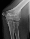
Figure 1
Initial fracture lateral X-ray depicting double arc sign. Olecranon has not yet fused.
Figure 2
Figure 2
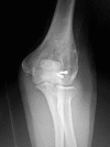
Initial fracture anteroposterior view X-ray. The fracture is not clearly evident in this view. Medial epicondyle has not yet fused.
Figure 3
Figure 3
CT scan three-dimensional reconstruction antepsoterior view depicting fractured capitellum and trochlea. Medial epicondylar fracture is also evident.
Figure 4
Figure 4
CT scan three-dimensional reconstruction lateral view depicting capitellum coronal shear as well as detachment of lateral epicondyle.
Figure 5
Figure 5
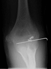
Two weeks postoperative X-ray anteposterior view.
Figure 6
Figure 6
Two weeks postoperative X-ray lateral view.
Figure 7
Figure 7
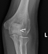
Six months postoperative X-ray anteroposterior view showing healing and no evidence of avascular necrosis. Medial epicondyle has fused.
Figure 8
Figure 8
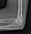
Six months postoperative X-ray lateral view showing healing and no evidence of avascular necrosis. Olecranon has fused.
Figure 9
Figure 9
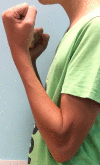
Patient review 6 months postoperative elbow extension.
Figure 10
Figure 10
Patient review 6 months postoperative elbow flexion.
Figure 11
Figure 11
Patient review 15 months postoperative X-ray anteroposterior view. No evidence of avascular necrosis.
Figure 12
Figure 12
Patient review 15 months postoperative X-ray lateral view. No evidence of avascular necrosis.

References

    1. Marion J, Faysse R. Fracture of the capitellum. Rev Chir Orthop 1962;48:484–90.
    1. McKee MD, Jupiter JB, Bamberger HB. Coronal shear fractures of the distal end of the humerus*. The J Bone & Jt Surg 1996;78:49–54. 10.2106/00004623-199601000-00007 - DOI - PubMed
    1. Singh AP, Singh AP, Vaishya R, et al. . Fractures of capitellum: a review of 14 cases treated by open reduction and internal fixation with Herbert screws. Int Orthop 2010;34:897–901. 10.1007/s00264-009-0896-9 - DOI - PMC - PubMed
    1. Ruchelsman DE, Tejwani NC, Kwon YW, et al. . Coronal plane partial articular fractures of the distal humerus: current concepts in management. J Am Acad Orthop Surg 2008;16:716–28. 10.5435/00124635-200812000-00004 - DOI - PubMed
    1. Dubberley JH, Faber KJ, Macdermid JC, et al. . Outcome after open reduction and internal fixation of capitellar and trochlear fractures. J Bone Joint Surg Am 2006;88:46–54. 10.2106/JBJS.D.02954 - DOI - PubMed

Publication types