. 2019 Aug 20;116(34):17039-17044.
doi: 10.1073/pnas.1905322116. Epub 2019 Aug 5.

Activity-evoked and spontaneous opening of synaptic fusion pores

Affiliations

Activity-evoked and spontaneous opening of synaptic fusion pores

Dinara Bulgari et al. Proc Natl Acad Sci U S A. .

Abstract

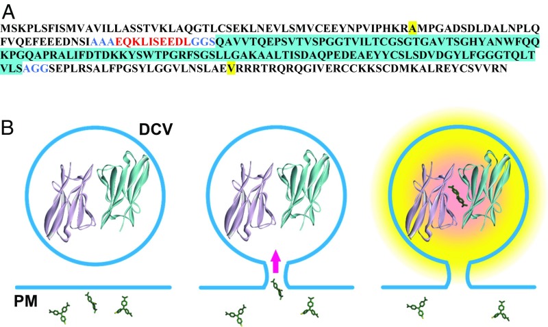
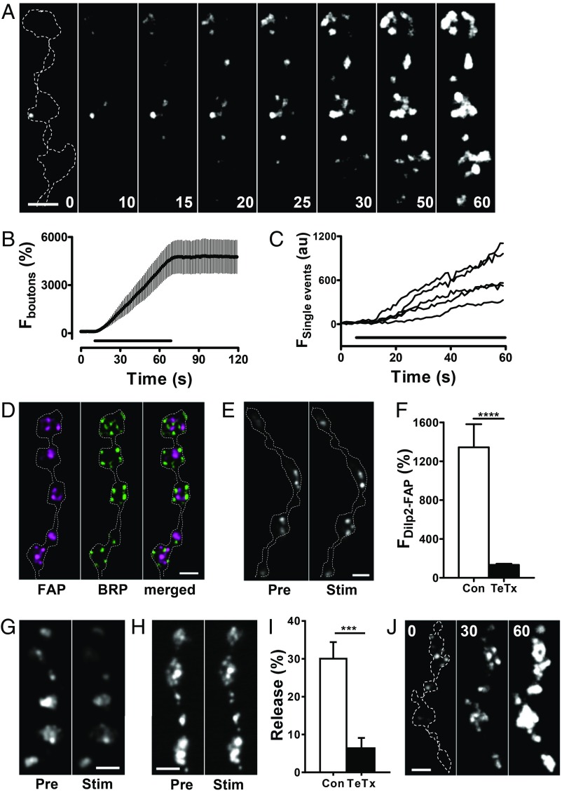
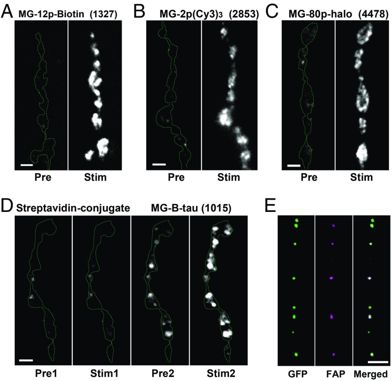
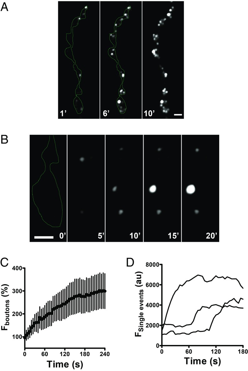
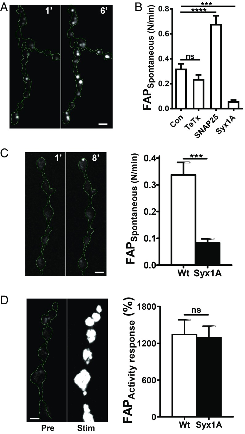
Synaptic release of neuropeptides packaged in dense-core vesicles (DCVs) regulates synapses, circuits, and behaviors including feeding, sleeping, and pain perception. Here, synaptic DCV fusion pore openings are imaged without interference from cotransmitting small synaptic vesicles (SSVs) with the use of a fluorogen-activating protein (FAP). Activity-evoked kiss and run exocytosis opens synaptic DCV fusion pores away from active zones that readily conduct molecules larger than most native neuropeptides (i.e., molecular weight [MW] up to, at least, 4.5 kDa). Remarkably, these synaptic fusion pores also open spontaneously in the absence of stimulation and extracellular Ca2+ SNARE perturbations demonstrate different mechanisms for activity-evoked and spontaneous fusion pore openings with the latter sharing features of spontaneous small molecule transmitter release by active zone-associated SSVs. Fusion pore opening at resting synapses provides a mechanism for activity-independent peptidergic transmission.

Keywords: Drosophila; fusion pore; neuromuscular junction; neuropeptide release; secretory granule.


Conflict of interest statement

The authors declare no conflict of interest.

Figures

Fig. 1.
Fig. 1.
Design of Dilp2-FAP. (A) Amino acid sequence of Dilp2-FAP. Dilp2 sequence is shown in plain black with the beginning and end of the C-peptide (from uniprot.org) highlighted in yellow. The c-Myc tag is red, linker sequences (AAA, GGS, and AGG) are blue, and the FAP monomer inserted in the C-peptide is black with blue highlighting. (B) Schematic of FAP experiments. Left, fusion with Dilp2 ensures that FAP will be highly concentrated in DCVs so that dimerization can occur (structure from 8). Extracellular MG molecules are shown at different orientations. Center, formation of a fusion pore with the plasma membrane (PM) allows extracellular membrane impermeant MG derivatives to enter the DCV lumen. Right, formation of complex between the nonfluorescent FAP dimer and MG derivative produces fluorescence. Note that small MG derivatives will diffuse through the fusion pore to increase fluorescence more quickly than exit of large MG-liganded FAP dimers and higher order MG-liganded Dilp2-FAP complexes.
Fig. 2.
Fig. 2.
Activity induces opening of DCV fusion pores in synaptic boutons. (A) Contrast-enhanced fluorescent mages of Dilp2-FAP expressing type Is boutons during 30 Hz stimulation for 60 s in the presence of MG-2p. Outline of boutons shown by the dashed line in the first panel. Numbers on the images indicate stimulation time in seconds. A low pass Gaussian blur filter was applied to images for presentation purposes. (B) Time course of bouton fluorescence increase during 30 Hz stimulation for 60 s in the presence of MG-2p. Data are from 4 animals (6 boutons). The bar indicates 30 Hz stimulation. (C) Representative traces of single puncta fluorescence increase during 30 Hz stimulation for 60 s. (D) Pseudocolor images of boutons coexpressing Dilp2-FAP (magenta) and BRP-GFP (green). (E) Representative images of boutons coexpressing Dilp2-FAP and a TeTx light chain before (Pre) and after (Stim) 70 Hz stimulation for 1 min in the presence of MG-2p. (F) Quantification of activity-dependent response in boutons expressing Dilp2-FAP in the control (Con) and boutons coexpressing Dilp2-FAP and TeTx as an increase in MG-2p activated FAP fluorescence in response to 70 Hz stimulation for 1 min. Data are from 6 animals (28 boutons) for each group. ****P < 0.001, unpaired t test. Quantification based on background subtracted unfiltered data. (G) Representative images of Dilp2-GFP expressing boutons before (Pre) and after 70 Hz stimulation for 1 min (Stim). (H) Representative images of boutons coexpressing Dilp2-GFP and TeTx Pre and Stim 70 Hz stimulation. (I) Quantification of activity-induced DCV-mediated release, measured as loss of GFP in the Con and TeTx expressing boutons. Data are from 5 Con animals and 6 TeTx animals. ***P < 0.001, unpaired t test. (J) Contrast-enhanced fluorescence images of Dilp2-FAP expressing boutons during 30 Hz stimulation for 60 s in the presence of MG-2p with Ba2+ instead of Ca2+ in HL3 saline. Outline of boutons shown by the dashed line in the first panel. Numbers on the images indicate stimulation time in seconds. Data are representative of results from 3 animals (Scale bars, 2 µm in all images.)
Fig. 3.
Fig. 3.
FAP labeling is mediated by fusion pores. Representative images of Dilp2-FAP expressing boutons before (Pre) and after 70 Hz stimulation for 1 min (Stim) in the presence of MG-12p-biotin (A), MG-(Cy3)3 (B), and MG-p80 (C). Boutons outlined with the dashed line. Numbers in parentheses are MW in daltons of the MG derivative. (D) Representative mages of Dilp2-FAP expressing boutons in the presence of MG-12p-biotin conjugated with streptavid before (Pre1) and after 70 Hz stimulation for 1 min (Stim1) and after replacing the conjugate with MG-B-tau (Pre2) and after 70 Hz stimulation for 1 min (Stim2). (E) Pseudocolor images of DCVs in the axon coexpressing Dilp2-FAP (magenta) and Dilp2-GFP (green) fixed in paraformaldehyde 5 min after 70 Hz stimulation for 60 s in the presence of MG-2p (Scale bars, 2 µm.)
Fig. 4.
Fig. 4.
Spontaneous fusion pore openings in Dilp2-FAP expressing boutons in the presence of MG-2p. (A) Representative images of Dilp2-FAP expressing boutons in the absence of Ca2+ after application of MG-2p. Numbers on the images indicate minutes. (B) Consecutive images of Dilp2-FAP expressing boutons in the absence of Ca2+after application of MG-2p show that individual puncta vary in whether they grow brighter with time. Numbers on the images indicate minutes (Scale bars, 2 µm.) (C) Time course of bouton fluorescence increase in the absence of Ca2+ after application of MG-2p. Data are from 5 animals (12 boutons). (D) Representative time courses of individual spontaneous fusion pore openings in the absence of Ca2+ after application of MG-2p.
Fig. 5.
Fig. 5.
SNARE dependence of spontaneous fusion pore opening. (A) Representative images of boutons coexpressing Dilp2-FAP and TeTx in the absence of Ca2+ after application of MG-2p. Numbers on the images indicate time in minutes. Note spontaneous brightening of the FAP signal. (B) Frequency of spontaneous fusion pore openings in the absence of Ca2+ analyzed as a number of fusion pore openings per bouton per minute. Data are from 6 animals for Con, (59 boutons), 4 animals for TeTx expressing boutons (19 boutons), 6 animals for SNAP25 G50E mutant (35 boutons), 7 animals for syntaxin 1A (Syx1A) RNAi expressing boutons (115 boutons). ***P < 0.001, ****P < 0.0001, Dunnett’s posttest following one-way ANOVA (P < 0.0001). (C, Left) Representative images of boutons coexpressing Dilp2-FAP and Syx1A RNAi after application to MG-2p in the presence of Ca2+. Numbers on the images represent minutes. (Right) Frequency of spontaneous fusion pore openings in normal Ca2+ saline. Data are from 4 animals in wild type (WT) animals (38 boutons) and 4 animals for Syx1A RNAi expressing boutons (43 boutons). ***P < 0.001, unpaired t test. (D, Left) Representative images of boutons coexpressing Dilp2-FAP and Syx1A RNAi Pre and Stim 70 Hz stimulation for 1 min. (Right) Quantification of Dilp2-FAP response in WT and Syx1A RNAi expressing boutons. Data are from 6 animals for WT (28 boutons) and 5 animals for Syx1A RNAi expressing boutons (28 boutons) (Scale bars, 2 μm.)

References

    1. Taghert P. H., Nitabach M. N., Peptide neuromodulation in invertebrate model systems. Neuron 76, 82–97 (2012). - PMC - PubMed
    1. Nusbaum M. P., Blitz D. M., Marder E., Functional consequences of neuropeptide and small-molecule co-transmission. Nat. Rev. Neurosci. 18, 389–403 (2017). - PMC - PubMed
    1. van den Pol A. N., Neuropeptide transmission in brain circuits. Neuron 76, 98–115 (2012). - PMC - PubMed
    1. Merighi A., Costorage of high molecular weight neurotransmitters in large dense core vesicles of mammalian neurons. Front. Cell. Neurosci. 12, 272 (2018). - PMC - PubMed
    1. Shin W., et al. , Visualization of membrane pore in live cells reveals a dynamic-pore theory governing fusion and endocytosis. Cell 173, 934–945.e12 (2018). - PMC - PubMed

Publication types

MeSH terms

LinkOut - more resources