Multicenter Study
. 2020 Mar;33(3):456-467.
doi: 10.1038/s41379-019-0334-5. Epub 2019 Aug 5.

Sclerosing epithelioid mesenchymal neoplasm of the pancreas - a proposed new entity

Affiliations
Multicenter Study

Sclerosing epithelioid mesenchymal neoplasm of the pancreas - a proposed new entity

Olca Basturk et al. Mod Pathol. 2020 Mar.

Abstract

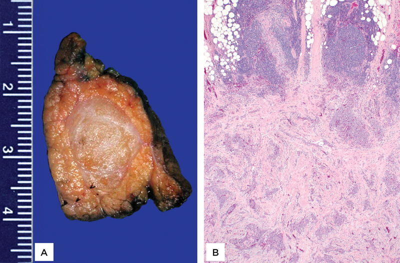
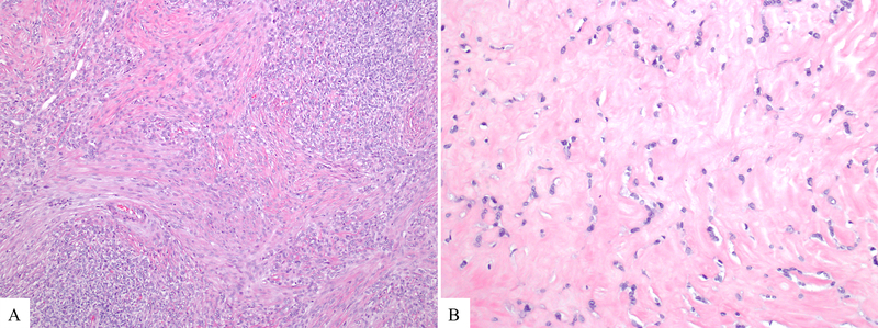
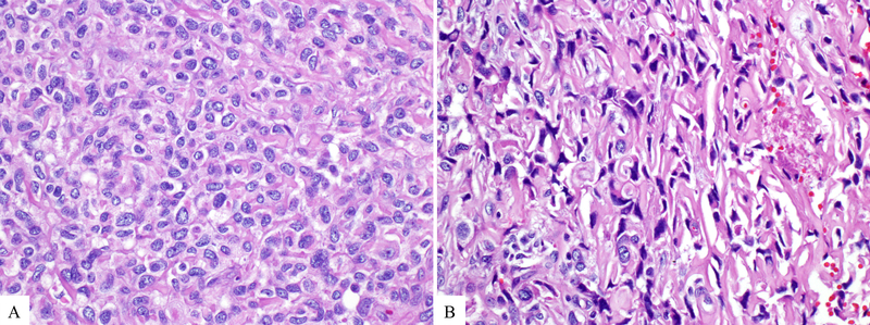
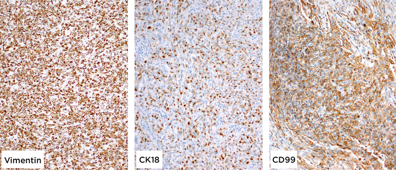
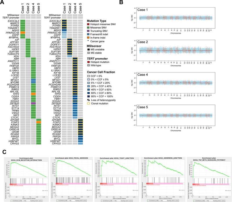
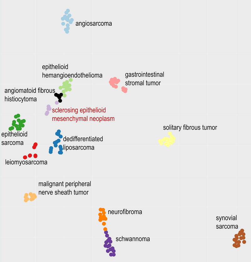
We have encountered pancreatic tumors with unique histologic features, which do not conform to any of the known tumors of the pancreas or other anatomical sites. We aimed to define their clinicopathologic features and whether they are characterized by recurrent molecular signatures. Eight cases were identified; studied histologically and by immunohistochemistry. Selected cases were also subjected to whole-exome sequencing (WES; n = 4), RNA-sequencing (n = 6), Archer FusionPlex assay (n = 5), methylation profiling using the Illumina MethylationEPIC (850k) array platform (n = 6), and TERT promoter sequencing (n = 5). Six neoplasms occurred in females. The mean age was 43 years (range: 26-75). Five occurred in the head/neck of the pancreas. All patients were treated surgically; none received neoadjuvant/adjuvant therapy. All patients are free of disease after 53 months of median follow-up (range: 8-94). The tumors were well-circumscribed, and the median size was 1.8 cm (range: 1.3-5.8). Microscopically, the unencapsulated tumors had a geographic pattern of epithelioid cell nests alternating with spindle cell fascicles. Some areas showed dense fibrosis, in which enmeshed tumor cells imparted a slit-like pattern. The predominant epithelioid cells had scant cytoplasm and round-oval nuclei with open chromatin. The spindle cells displayed irregular, hyperchromatic nuclei. Mitoses were rare. No lymph node metastases were identified. All tumors were positive for vimentin, CD99 and cytokeratin (patchy), while negative for markers of solid pseudopapillary neoplasm, neuroendocrine, acinar, myogenic/rhabdoid, vascular, melanocytic, or lymphoid differentiation, gastrointestinal stromal tumor as well as MUC4. Whole-exome sequencing revealed no recurrent somatic mutations or amplifications/homozygous deletions in any known oncogenes or tumor suppressor genes. RNA-sequencing and the Archer FusionPlex assay did not detect any recurrent likely pathogenic gene fusions. Single sample gene set enrichment analysis revealed that these tumors display a likely mesenchymal transcriptomic program. Unsupervised analysis (t-SNE) of their methylation profiles against a set of different mesenchymal neoplasms demonstrated a distinct methylation pattern. Here, we describe pancreatic neoplasms with unique morphologic/immunophenotypic features and a distinct methylation pattern, along with a lack of abnormalities in any of key genetic drivers, supporting that these neoplasms represent a novel entity with an indolent clinical course. Given their mesenchymal transcriptomic features, we propose the designation of "sclerosing epithelioid mesenchymal neoplasm" of the pancreas.


Figures

Figure 1:
Figure 1:
The tumors were well-circumscribed and solid. Cut surfaces were tan-white with firm/ sclerotic consistency (A). Sections revealed epithelioid and spindled cell neoplasms associated with extensive fibrosis as well as dense lymphoid aggregates at the periphery (B).
Figure 2.
Figure 2.
The density of tumor cells varied significantly among the tumors and throughout each tumor. Although in some areas the tumor cells formed cellular sheets (A); other areas showed dense hyaline fibrosis with only rare nests of neoplastic cells (B).
Figure 3:
Figure 3:
The tumor cells exhibited variable morphology. Most cells were epithelioid and contained scant cytoplasm and round to oval nuclei with open chromatin, some with conspicuous nucleoli (A). Spindled cells displayed more irregular, hyperchromatic nuclei (B).
Figure 4:
Figure 4:
All tumors were positive for vimentin, CD99 and cytokeratins (AE1:AE3 and CK18, both patchy), while negative for markers of solid pseudopapillary neoplasm, acinar- myogenic/rhabdoid-, vascular-, melanocytic-, lymphocytic differentiation as well as MUC4.
Figure 5.
Figure 5.. Genomic and transcriptomic characterization of sclerosing epithelioid mesenchymal neoplasm of the pancreas.
A) Non-synonymous somatic mutations in sclerosing epithelioid mesenchymal neoplasm of the pancreas detected by whole-exome sequencing. The mutation types (left) and cancer cell fractions of each mutation (right) are shown, color-coded according to the legend. The phenobar (top) provides information about the presence of TERT promoter hotspot mutations and the microsatellite instability sensor score (microsatellite instability). Indel, small insertion and deletion; MS, microsatellite; SNV, single nucleotide variant. B) Chromosome plots of the four sclerosing epithelioid mesenchymal neoplasm of the pancreas subjected to whole-exome sequencing. Log2-ratios plotted on the y‐axis according to their genomic coordinates on the x‐axis. C) Results of the Gene Set Enrichment Analysis of genes overexpressed in six sclerosing epithelioid mesenchymal neoplasm of the pancreas subjected to RNA-sequencing. The pathways found to be enriched are shown in the title of each plot.
Figure 6.
Figure 6.
Unsupervised analysis (t-SNE dimensionality reduction algorithm) of the most variable 20,000 CpG sites demonstrates a distinct clustering of these pancreatic neoplasms as compared to other mesenchymal neoplasms in the differential diagnosis.

References

    1. Pareja F, Lee JY, Brown DN, Piscuoglio S, Gularte-Merida R, Selenica P, et al. The genomic landscape of mucinous breast cancer. J Natl Cancer Inst 2019. - PMC - PubMed
    1. Ashley CW, Da Cruz Paula A, Kumar R, Mandelker D, Pei X, Riaz N, et al. Analysis of mutational signatures in primary and metastatic endometrial cancer reveals distinct patterns of DNA repair defects and shifts during tumor progression. Gynecol Oncol 2018. - PMC - PubMed
    1. Li H, Durbin R. Fast and accurate short read alignment with Burrows-Wheeler transform. Bioinformatics 2009; 25:1754–60. - PMC - PubMed
    1. Mckenna A, Hanna M, Banks E, Sivachenko A, Cibulskis K, Kernytsky A, et al. The Genome Analysis Toolkit: a MapReduce framework for analyzing next-generation DNA sequencing data. Genome Res 2010; 20:1297–303. - PMC - PubMed
    1. Cibulskis K, Lawrence MS, Carter SL, Sivachenko A, Jaffe D, Sougnez C, et al. Sensitive detection of somatic point mutations in impure and heterogeneous cancer samples. Nat Biotechnol 2013; 31:213–9. - PMC - PubMed

Publication types

MeSH terms

Substances