. 2019 Sep;3(9):706-716.
doi: 10.1038/s41551-019-0434-z. Epub 2019 Aug 5.

Sustained delivery and molecular targeting of a therapeutic monoclonal antibody to metastases in the central nervous system of mice

Affiliations

Sustained delivery and molecular targeting of a therapeutic monoclonal antibody to metastases in the central nervous system of mice

Jing Wen et al. Nat Biomed Eng. 2019 Sep.

Abstract

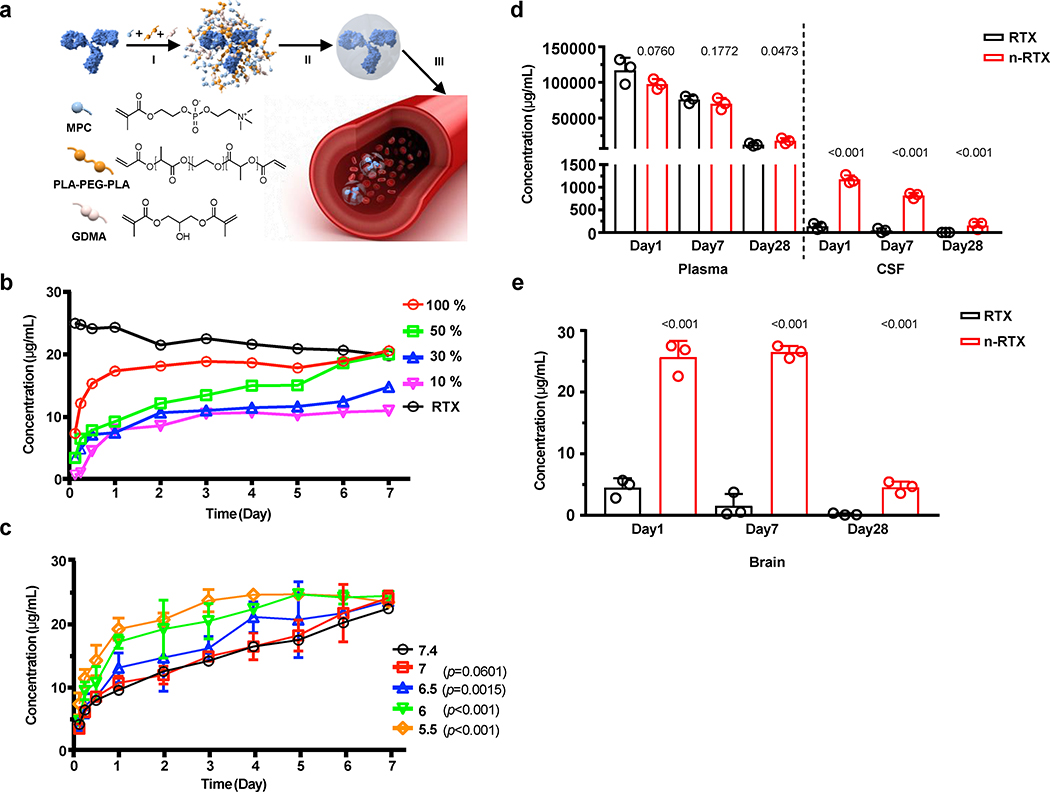
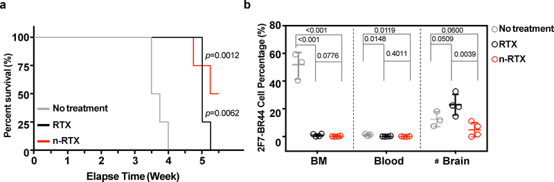
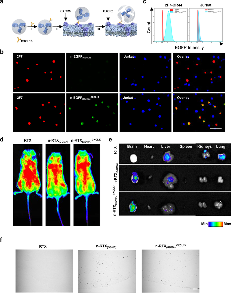
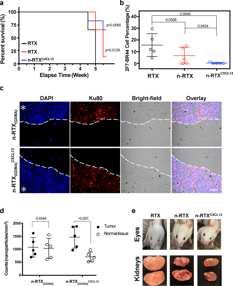
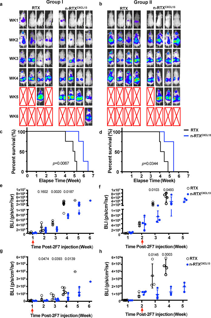
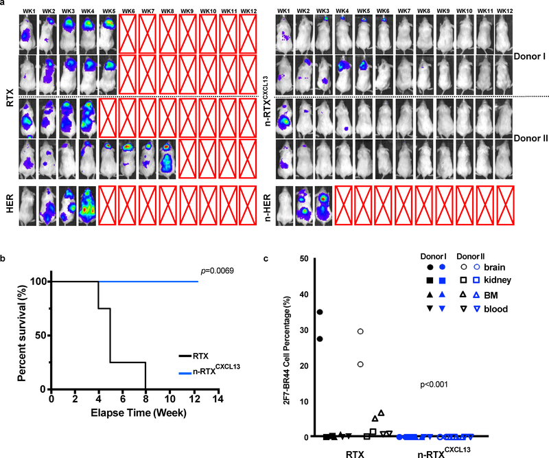
Approximately 15-40% of all cancers develop metastases in the central nervous system (CNS), yet few therapeutic options exist to treat them. Cancer therapies based on monoclonal antibodies are widely successful, yet have limited efficacy against CNS metastases, owing to the low levels of the drug reaching the tumour site. Here, we show that the encapsulation of rituximab within a crosslinked zwitterionic polymer layer leads to the sustained release of rituximab as the crosslinkers are gradually hydrolysed, enhancing the CNS levels of the antibody by approximately tenfold with respect to the administration of naked rituximab. When the nanocapsules were functionalized with CXCL13-the ligand for the chemokine receptor CXCR5, which is frequently found on B-cell lymphoma-a single dose led to improved control of CXCR5-expressing metastases in a murine xenograft model of non-Hodgkin lymphoma, and eliminated lymphoma in a xenografted humanized bone marrow-liver-thymus mouse model. Encapsulation and molecular targeting of therapeutic antibodies could become an option for the treatment of cancers with CNS metastases.


Conflict of interest statement

Competing interests

I.S.Y.C has a financial interest in CSL Behring. The remaining authors declare no competing interests.

Figures

Fig. 1 |
Fig. 1 |. Timed-release nanocapsules facilitate brain delivery of RTX in mice.
a, Scheme of the synthesis and release of timed-release RTX nanocapsules by (I) enriching MPC monomer and hydrolysable crosslinker PLA-PEG-PLA and slowly hydrolysable crosslinker GDMA around a RTX molecule, (II) in situ polymerization of the monomer and crosslinkers forming a thin shell of polymer around a RTX molecule, and (III) releasing RTX when polymer shells are degraded under physiological conditions by hydrolysis of the crosslinkers. b, RTX nanocapsules were synthesized with mixed crosslinkers GDMA and PLA-PEG-PLA, at which the ratios of PLA-PEG-PLA were 100%, 50%, 30%, and 10%. Twenty-five μg of each nanoencapsulated RTX or native RTX (RTX) was added into 1mL of PBS and incubated at 37 °C for 1 week. The concentration of free RTX was measured by ELISA. c, The release rates of RTX nanocapsules with 50% PLA-PEG-PLA crosslinkers were compared in PBS buffers with various pH values presenting physiological conditions (pH=7.4 and 7) and acidic pH conditions (pH=6.5, 6, and 5.5). Twenty-five μg of each sample were added into 1mL of PBS buffer and incubated at 37 °C for up to 1 week. The concentration of free RTX was measured by ELISA. Data are shown as means ± standard deviations (SDs) of biological duplicates. The statistical significance of the pH7.4 (physiological condition) was assessed by p-value, calculated by one-tailed paired t-test with Welch’s correction. d,e, B6 mice (n=3) were administrated with 20 mg/kg of native RTX or RTX nanocapsules with 50% PLA-PEG-PLA crosslinkers (n-RTX) via retro-orbital vein. Plasma, CSF (b), and perfused brain tissue samples (c) were collected and concentrations of free RTX were measured by ELISA on days 1, 7, and 28. Data are shown as means ± SDs of biological triplicates. The statistical significance of the data for the native RTX group was assessed via p-values (provided), calculated by one-tailed unpaired t-test with Welch’s correction.
Fig. 2 |
Fig. 2 |. Nanocapsulation enhances anti-lymphoma activity of RTX against CNS lymphomas in the 2F7-BR44 murine xenograft model.
2F7-BR44 cells (2×10⁶/animal) in 100 μL Hank’s balanced salt solution were injected into NSG mice via tail vein. Five days after injection, mice were randomly separated into three groups: no treatment (n=3), native RTX and n-RTX treatment (n=4). Native RTX or n-RTX was injected via retro-orbital vein at 4 mg/kg/day for 5 days. a, Kaplan-Meier survival curves of the mice in each group were plotted relative to the number of weeks after 2F7-BR44 cell injection. The statistical significance was shown with a p-value calculated by the log-rank test. b, Tissue distribution of 2F7-BR44 cells in blood, perfused bone marrow (BM) and brain was analyzed by flow cytometry at endpoints. Data are shown means ± SDs of biological triplicates (No treatment) or quadruplicates (RTX or n-RTX). Statistical significance was calculated with respect to the no treatment group using a p-value calculated by one-tailed unpaired t-test with Welch’s correction. #: Note that the apparent higher tumor burden in brain of the native RTX group compared to the no-treatment group is due to one-week longer duration of the native RTX group versus the no-treatment group which were euthanized one week earlier due to massive systemic tumor growth.
Fig. 3 |
Fig. 3 |. CXCL13 conjugation mediates 2F7-BR44 cell specific nanocapsule targeting without affecting overall biodistribution.
a, Scheme of CXCL13 conjugation on n-RTX(GDMA). CXCL13 was conjugated to n-RTX(GDMA) at a molar ratio of 1:1 through copper-free click chemistry. b,c, EGFP nanocapsules with 100% slowly hydrolysable crosslinkers, GDMA, (n-EGFP(GDMA)) conjugated with or without CXCL13 (0.1 mg/ml each) were incubated 1 hour at 37⁰C with a mixed cell population of 2F7-BR44 cells (2F7, mStrawberry+) and Jurkat T cells (Jurkat, BFP+). The binding of each nanocapsule on each cell population was analyzed on Leica DMi8 inverted microscope (b) or on BD LSRFortessa (c). The same experiments were repeated independently three times. d,e, NSG mice (n=3) were administrated with 20 mg/kg of carboxytetramethylrhodamine (TAMRA)-labelled RTX, n-RTX(GDMA) and n-RTX(GDMA)CXCL13 via tail vein. Fluorescence images of whole body (d) and each organ (e) were taken 1 day after administration. f, NSG mice (n=3) were administrated with 20 mg/kg of gold-nanoparticle labelled RTX, n-RTX(GDMA) and n-RTX(GDMA)CXCL13 via tail vein. Brain tissues were obtained 1 day after administration and processed for immunohistochemical analysis. Gold nanoparticles in paraffin sections were analyzed following silver enhancement, in which present as black depositions. Bar=50 μm.
Fig. 4 |
Fig. 4 |. CXCL13 conjugation improves anti-lymphoma activity of n-RTX.
a,b, 2F7-BR44 cells were injected to NSG mice via tail vein (2×10⁶/animal). Five days after the injection, mice were randomly separated into three groups (n=5): native RTX, n-RTX, and n-RTXCXCL13. Each form of RTX was administrated via retro-orbital injection at 4mg/kg/day for 5 days. Kaplan-Meier survival curves of the mice in each group were plotted relative to the number of weeks after 2F7-BR44 cell injection (a). Statistical significance was shown with a p-value calculated by the log-rank test. Percentages of 2F7-BR44 cells in the brain were analyzed by flow cytometry at endpoints following cardiac perfusion (b). The statistical significance was calculated to the native RTX group using a p-value calculated by one-tailed unpaired t-test with Welch’s correction. Data are shown means ± SDs of biological quintuplicates. c,d, Localizations of gold-labelled n-RTX(GDMA) with or without CXCL13 conjugation were analyzed in the brain of 2F7-BR44 xenograft NSG mice (n=3) 3 weeks after xenograft. One day after administration of gold-labelled n-RTX(GDMA) or n-RTX(GDMA)CXCL13 via retro-orbital injection, brain tissue samples were processed for immunohistochemical analysis (c). Gold nanoparticles in paraffin sections were analyzed following silver enhancement, presented as black depositions. 2F7-BR44 cells were stained with anti-human Ku80 antibody and rhodamine red-X anti-rabbit IgG. The nuclei were stained by 4′,6-diamidino-2-phenylindole (DAPI). Asterisks indicate the tumor site while the white dash curves illustrate the boundary of tumor-normal parenchyma. Bar=25 μm. The average counts of gold-labelled n-RTX(GDMA) or n-RTX(GDMA)CXCL13 per mm2 from 5 spots in representatives in Fig.4C (total counts from samples administrated with n-RTX(GDMA) or n-RTX(GDMA)CXCL13 are approximately 11,000, respectively) in both the tumor site and normal tissue site were quantified by ImageJ (d). The statistical significance was calculated to normal tissue using a p-value calculated by one-tailed unpaired t-test with Welch’s correction. e, Representative images of intraocular lymphoma and lymphoma surrounding the kidney in each group.
Fig. 5 |
Fig. 5 |. CXCL13 conjugation improves anti-lymphoma activity of n-RTX at different stages of lymphoma progression.
CXCL13 was conjugated to n-RTX at a molar ratio of 1:1 through copper-free click chemistry (n-RTXCXCL13). 2F7-BR44 cells expressing firefly luciferase (2F7-BR44-Luc) were injected to NSG mice via tail vein (2×10⁶/animal) (a-h). Mice were randomly separated into four groups (n=4): native RTX or n-RTXCXCL13 treatment at week 1 (a,c,e,g, Group I) or week 2 (b,d,f,h, Group II) after 2F7-BR44-Luc cell injection. Each form of RTX was injected via retro-orbital vein at 4 mg/kg/day for 5 days. a,b, The tumor progression and metastasis status were monitored weekly by using bioluminescence imaging on IVIS Lumina II In Vivo Imaging system. Red arrows show the treatment time in each figure. Expansion and metastasis status of 2F7-BR44-Luc cells in NSG mice treated with native RTX or n-RTXCXCL13 at week 1 (Group I) or week 2 (Group II) as visualized by bioluminescence imaging. The red X boxes represent deceased mice. Sensitivity settings were adjusted at each time point to maintain 500–5000 counts per pixel and assigned the same color scale for both groups. c,d, Kaplan-Meier survival curves of the mice in each group were plotted relative to the number of weeks after 2F7-BR44-Luc cell injection. e,f, The bioluminescence intensity (BLI) in the whole body was compared between mice with treatment by native RTX and n-RTXCXCL13 in both groups and expressed in photons per second per square centimeter per steradian (p/s/cm2/sr). g,h, The BLI from head area was compared between mice with treatment by native RTX and n-RTXCXCL13 in both groups. Data are shown means ± SDs of biological quadruplicates. Statistical significance based on the native RTX group using a p-value was calculated by one-tailed unpaired t-test with Welch’s correction.
Fig. 6 |
Fig. 6 |. n-RTXCXCL13 exhibits superior anti-lymphoma activity against CNS lymphomas in the 2F7-BR44-Luc cell xenograft humanized mice.
2F7-BR44-Luc cells (2×10⁶/animal) were injected into humanized BLT mice generated from two independent donors (Donors I and II) via tail vein (a-c). Mice were randomly separated into two groups for treatment by native RTX or n-RTXCXCL13 at week 1 after the injection. Each form of RTX was administrated via retro-orbital vein at 4 mg/kg/day for 5 days. The tumor progression status was tracked weekly using bioluminescence imaging on IVIS Lumina II In Vivo Imaging system. a, Biodistribution of 2F7-BR44-Luc cells in humanized mice treated with native RTX or n-RTXCXCL13 at week 1 as visualized by luciferase bioimaging. The red X boxes represent deceased mice. Sensitivity settings were adjusted at each time point to maintain 250–5000 counts per pixel and assigned the same color scale for all treatment groups. b, Kaplan-Meier survival curves of the mice were plotted relative to the number of weeks after injection. Necropsies were performed at the end of the experiment (week 13). The statistical significance was shown with a p-value calculated by the log-rank test. c, Percentages of 2F7-BR44-Luc cells in tissues were assessed by flow cytometry following cardiac perfusion at the endpoints. HER: naive Herceptin. n-HER: Herceptin nanocapsule. The statistical significance was calculated for all tissues from the native RTX group using a p-value, calculated by one-tailed unpaired t-test with Welch’s correction.

References

    1. Chambers AF, Groom AC & MacDonald IC Dissemination and growth of cancer cells in metastatic sites. Nat Rev Cancer 2, 563–572 (2002). - PubMed
    1. Aragon-Ching JB & Zujewski JA CNS metastasis: an old problem in a new guise. Clin Cancer Res 13, 1644–1647 (2007). - PubMed
    1. Tosoni A, Ermani M & Brandes AA The pathogenesis and treatment of brain metastases: a comprehensive review. Crit Rev Oncol Hematol 52, 199–215 (2004). - PubMed
    1. Rubenstein JL, et al. Rituximab therapy for CNS lymphomas: targeting the leptomeningeal compartment. Blood 101, 466–468 (2003). - PubMed
    1. Zhang Y & Pardridge WM Mediated efflux of IgG molecules from brain to blood across the blood-brain barrier. J Neuroimmunol 114, 168–172 (2001). - PubMed

Publication types