

Paracentral acute middle maculopathy as a cause of unexplained visual loss in central retinal vein occlusion
- PMID: 31384162
- PMCID: PMC6664399
- DOI: 10.1016/j.sjopt.2018.07.005
Paracentral acute middle maculopathy as a cause of unexplained visual loss in central retinal vein occlusion
Abstract
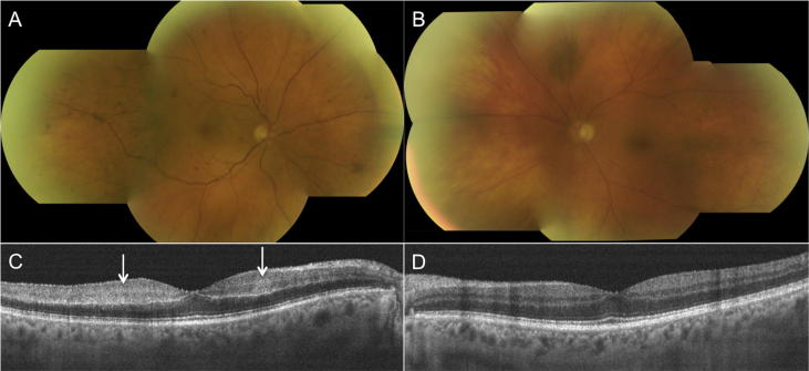
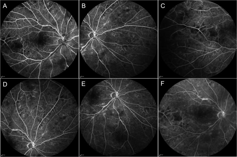
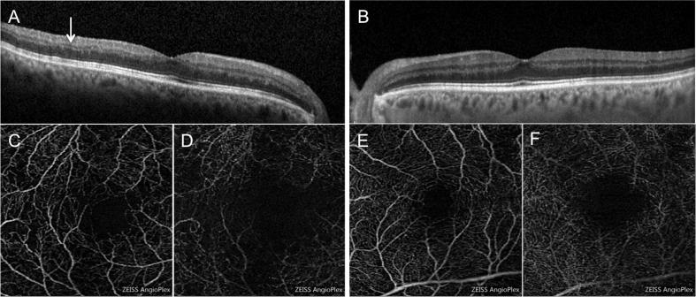
A 79-year-old man presented with unilateral unexplained sudden onset visual loss in the setting of central retinal vein occlusion (CRVO). Non ischemic CRVO in the right eye (RE) was confirmed on fluorescein angiography. Spectral domain optical coherence tomography (SD-OCT) showed absence of macular edema and hyperreflective band-like lesions in the middle retinal layers of the RE suggesting a diagnosis of paracentral acute middle maculopathy (PAMM). Patient was observed and after 3 months, best-corrected visual acuity in the RE spontaneously improved from 38 to 56 ETDRS letters. SD-OCT scans showed thinning of the inner nuclear layer of the RE. OCT angiography in the RE revealed a mild attenuation of the vascular flow signal in the superficial capillary plexus and patchy areas of vascular flow void in the deep capillary plexus, as compared to the fellow eye. The present case outlines the importance of recognising PAMM as a cause of unexplained visual loss. In the setting of a CRVO with sudden vision loss and absence of macular edema, clinicians should pay attention to any hyperreflectivity and/or to thinning of the middle retinal layers on SD-OCT.
Keywords: Central retinal vein occlusion; Deep capillary ischemia; Optical coherence tomography angiography; Paracentral acute middle maculopathy; spectral domain optical coherence tomography.
Figures
References
-
- Sarraf D., Rahimy E., Fawzi A.A. Paracentral acute middle maculopathy: a new variant of acute macular neuroretinopathy associated with retinal capillary ischemia. JAMA Ophthalmol. 2013;131:1275–1287. - PubMed
-
- Rahimy E., Kuehlewein L., Sadda S.R., Sarraf D. Paracentral acute middle maculopathy: what we knew then and what we know now. Retina. 2015;35:1921–1930. - PubMed
-
- Chen X., Rahimy E., Sergott R.C. Spectrum of retinal vascular diseases associated with paracentral acute middle maculopathy. Am J Ophthalmol. 2015;160:26–34. - PubMed
-
- Rahimy E., Sarraf D., Dollin M.L., Pitcher J.D., Ho A.C. Paracentral acute middle maculopathy in non ischemic central retinal vein occlusion. Am J Ophthalmol. 2014;158:372–380. - PubMed
-
- Spaide R.F., Klancnik J.M., Cooney M.J. Retinal vascular layers imaged by fluorescein angiography and optical coherence tomography angiography. JAMA Ophthalmol. 2015;133:45–50. - PubMed
LinkOut - more resources
Full Text Sources
