Case Reports
. 2019 May 24;32(3):382-384.
doi: 10.1080/08998280.2019.1596502. eCollection 2019 Jul.

Percutaneous transorbital embolization of a carotid cavernous fistula

Affiliations
Case Reports

Percutaneous transorbital embolization of a carotid cavernous fistula

Lance J Lyons et al. Proc (Bayl Univ Med Cent). .

Abstract

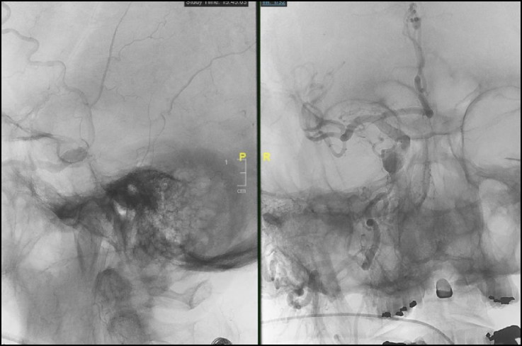
This case report highlights utilization of image-guided, percutaneous transorbital direct cavernous sinus puncture to embolize an anteriorly draining carotid cavernous fistula (CCF) when conventional transarterial and transvenous approaches were not feasible. An 86-year-old man with a known posterior draining CCF developed acute unilateral proptosis, pain, and vision loss ("red-eyed shunt"). Cerebral angiogram revealed the dural CCF to be draining anteriorly into partially thrombosed ophthalmic veins. After failed transarterial and transvenous attempts, a percutaneous transorbital approach was used to successfully embolize the fistula using the Onyx Liquid Embolic System according to the visual needle path generated by the Seimens Syngo iGuide. To our knowledge, this is the first reported case of percutaneous transorbital direct embolization of a CCF utilizing the Seimens Syngo iGuide.

Keywords: Carotid cavernous fistula; Seimens Syngo iGuide; onyx liquid embolic system; percutaneous transorbital direct cavernous sinus puncture.


Figures

Figure 1.
Figure 1.
Intraoperative angiogram, transvenous attempt. Utilizing a direct percutaneous approach, two needles were used to try to select the inferior ophthalmic vein but were unsuccessful. Subsequently, a stiff micropuncture set was used to select the most anterior aspect of the left cavernous sinus.
Figure 2.
Figure 2.
Intraoperative angiogram, fistula embolization. Multiple coils and Onyx 34 embolization material were used to obliterate the fistula.

References

    1. Hawke SH, Mullie MA, Hoyt WF, Hallinan JM, Halmagyi GM. Painful oculomotor nerve palsy due to dural-cavernous sinus shunt. Arch Neurol. 1989;46:1252–1255. doi: 10.1001/archneur.1989.00520470126040. - DOI - PubMed
    1. Thinda S, Melson MR, Kuchtey RW. Worsening angle closure glaucoma and choroidal detachments subsequent to closure of a carotid cavernous fistula. BMC Ophthalmol. 2012;12:12–28. - PMC - PubMed
    1. Sia PI, Sia DIT, Scroop R, Selva D. Orbital compartment syndrome following transvenous embolization of carotid-cavernous fistula. Orbit. 2014;33:52–54. doi: 10.3109/01676830.2013.841717. - DOI - PubMed
    1. Chaudhry IA, Elkhamry SM, Al-Rashed W, Bosley TM. Carotid cavernous fistula: opthalmological implications. Middle East Afr J Ophthalmol. 2009;16:57–63. doi: 10.4103/0974-9233.53862. - DOI - PMC - PubMed

Publication types

LinkOut - more resources