

Immunohistochemical study of psoriatic plaques and perilesional skin in psoriasis vulgaris patients: A pilot study
- PMID: 31384319
- PMCID: PMC6639978
- DOI: 10.3892/etm.2019.7596
Immunohistochemical study of psoriatic plaques and perilesional skin in psoriasis vulgaris patients: A pilot study
Abstract
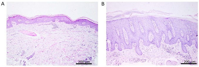
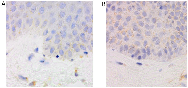
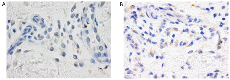
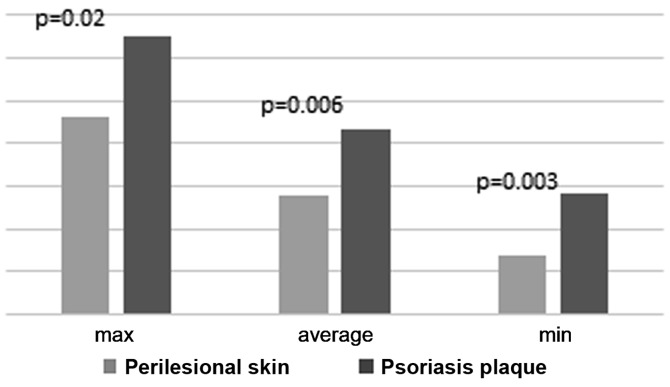
Psoriasis vulgaris, a chronic inflammatory skin disorder, is the result of immune mediated processes, genetic background and environmental factors. Prolactin and the vascular endothelial growth factor seem to play a key role in psoriasis pathogenesis regarding hyperproliferation of epidermal keratinocytes and dermal vascular ectasia. The aim of the study was to investigate the expression of tumor necrosis factor-α (TNF-α), vascular endothelial growth factor receptor 2 (VEGFR2) and prolactin receptor (PRLR) in psoriatic skin by immunohistochemical analysis and to evaluate the correlation with disease severity. Two skin biopsies, psoriatic lesion and perilesional skin, obtained by punch biopsy from 19 nontreated psoriasis patients were examined in hematoxylin and eosin staining and immunohistochemistry (IHC) for TNF-α, VEGFR2 and PRLR. The indirect IHC reaction was carried out automatically and visualized by 3,3-diaminobenzidine (DAB) technique. The average number of DAB-positive cells and the intensity of cell staining were quantified on a predefined scale. The results show a significant difference in the quantity and distribution of TNF-α positive cells in the two sample groups. In psoriatic plaque skin, an increased expression of TNF-α was found in the perivascular dermis and epidermic keratinocytes. In perilesional skin the immunostaining was predominant in the basal layer keratinocytes, while in psoriatic plaque, all the layers were positively marked, with stronger expression at the base. A statistically significant difference was found between the intensity of the immunostaining in the two types of tissue. Positive cells for VEGFR2 and PRL were identified in the basal layer keratinocyte cells (VEGFR2), sweat glands and hair outer shaft sheath (PRLR), without significant differences between the two types of samples. Our findings confirm the importance of TNF-α in psoriasis pathogenesis and a positive correlation with lesions severity. No significant differences were found for VEGFR2 and PRLR, but additional studies are necessary to establish their role.
Keywords: prolactin; psoriasis-immunohistochemistry-TNF-α; vascular endothelial growth factor.
Figures

References
-
- Boda D, Negrei C, Nicolescu F, Balalau C. Assessment of some oxidative stress parameters in methotrexate treated psoriasis patients. Farmacia. 2014;62:704–710.
-
- Raţiu MP, Purcărea I, Popa F, Purcărea VL, Purcărea TV, Lupuleasa D, Boda D. Escaping the economic turn down through performing employees, creative leaders and growth driver capabilities in the Romanian pharmaceutical industry. Farmacia. 2011;59:119–130.
LinkOut - more resources
Full Text Sources