Review
. 2019 Apr-Jun;15(2):99-104.
doi: 10.14797/mdcj-15-2-99.

Advanced Cardiac Imaging for Complex Adult Congenital Heart Diseases

Affiliations
Review

Advanced Cardiac Imaging for Complex Adult Congenital Heart Diseases

Maan Malahfji et al. Methodist Debakey Cardiovasc J. 2019 Apr-Jun.

Abstract

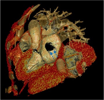
The population of patients with adult congenital heart disease has grown and is currently estimated to include approximately 1 million people in the United States. Cardiologists and imagers frequently encounter complex patients who have undergone multiple prior operations and interventions. A myriad of imaging tests are currently available, including echocardiography, cardiovascular magnetic resonance imaging, and computed tomography, all of which collectively provide invaluable information on cardiac anatomy and hemodynamics. Advanced imaging plays a role in diagnosis and preprocedural planning and also determines the need and frequency of follow-up. This article provides a contemporary review of the current role of cardiac imaging in patients with complex congenital heart disease.

Keywords: adult congenital heart disease; cardiac computed tomography; cardiac magnetic resonance imaging; three-dimensional contrast-enhanced angiography.


Conflict of interest statement

Conflict of Interest Disclosure: The authors have completed and submitted the Methodist DeBakey Cardiovascular Journal Conflict of Interest Statement and none were reported.

Figures

Figure 1.
Figure 1.
Two-chamber sagittal view on cardiac computed tomography showing septal ridge separating the left atrium into two chambers, one draining the pulmonary veins and the other one connecting to the mitral inflow, consistent with Cor triatriatum sinister.
Figure 2.
Figure 2.
(A) Cardiac computed tomography axial image and (B) volume-rendered 3-dimensional image showing anomalous origin of left main from the right coronary cusp. There is no evidence of malignant features including lack of intera-arterial or intramural course.
Figure 3.
Figure 3.
Cardiac computed tomography, coronalimage showing extensive mediastinal and peribronchial collateral circulation in a patient with coronary artery to pulmonary artery fistula.
Figure 4.
Figure 4.
Volume-rendered imaging with cardiac computed tomography and 3-dimensional depiction of secundum atrial septal defect (arrows).
Figure 5.
Figure 5.
Three-chamber sagittal view on cardiac computed tomography showing a patient with D-transposition of the great arteries after arterial switch surgery. The morphologic right ventricle is connected to the aorta, hence it is the systemic ventricle. The pulmonary veins were baffled to the right atrium (asterisk).
Figure 6.
Figure 6.
(A, B) Steady-state free precession (SSFP) cardiac magnetic resonance imaging (CMR) sequence, 4-chamber view, showing large secundum atrial septal defect (ASD, asterisk, 2.0 × 1.5 cm). (B) Corresponding phase-contrast imaging at same plane showing left-to-right shunting across the defect. (C, D) SSFP CMR sequence, sagittal view, again showing ASD with corresponding phase-contrast imaging at same plane. (D) There was a dilated right ventricle (RV), with an RV end-diastolic volume of 261 mL, and normal RV systolic function, with an RV ejection fraction of 52%. The net Qp:Qs in this case was 1.5:1.
Figure 7.
Figure 7.
A patient with repaired tetralogy of Fallot/pulmonic stenosis with pulmonic valvotomy and transannular patch presented with worsening shortness of breath. (A) Cine steady-state free precession (SSFP) computed magnetic resonance imaging (CMR) sequence 4-chamber view showing severe right ventricular (RV) enlargement (RV end diastolic volume of 165 mL/m2). (B) Cine SSFP CMR sequence sagittal oblique view at the pulmonic valve level showing transannular patch. (C) Phase-contrast CMR imaging at the same location as (B) in diastole showing pulmonic regurgitation (quantitatively, the regurgitant volume was 132 mL and regurgitant fraction was 78%).
Figure 8.
Figure 8.
Sagittal view, 3-dimensional magnetic resonance angiography of the aorta showing severe coarctation (A, arrows) with post-stenotic dilation of the descending aorta. In addition, there is extensive collateral circulation in the chest originating from the descending thoracic aorta (B, arrows).

References

    1. Burchill LJ, Huang J, Tretter JT et al. Noninvasive Imaging in Adult Congenital Heart Disease. Circ Res. 2017 Mar 17;120(6):995–1014. - PubMed
    1. Dacher JN, Barre E, Durand I et al. CT and MR imaging in congenital cardiac malformations: Where do we come from and where are we going? Diagn Interv Imaging. 2016 May;97(5):505–12. - PubMed
    1. Bhat V, Belaval V, Gadabanahalli K, Raj V, Shah S. Illustrated Imaging Essay on Congenital Heart Diseases: Multimodality Approach Part I: Clinical Perspective, Anatomy and Imaging Techniques. J Clin Diagn Res. 2016 May;10(5):TE01-6. - PMC - PubMed
    1. D'Alto M, Dimopoulos K, Budts W et al. Multimodality imaging in congenital heart disease-related pulmonary arterial hypertension. Heart. 2016 Jun 15;102(12):910–8. - PubMed
    1. Han BK, Rigsby CK, Hlavacek A et al. Computed Tomography Imaging in Patients with Congenital Heart Disease Part I: Rationale and Utility. An Expert Consensus Document of the Society of Cardiovascular Computed Tomography (SCCT): Endorsed by the Society of Pediatric Radiology (SPR) and the North American Society of Cardiac Imaging (NASCI) J Cardiovasc Comput Tomogr. 2015 Nov-Dec;9(6):475–92. Society of Cardiovascular Computed Tomography; Society of Pediatric Radiology; North American Society of Cardiac Imaging. - PubMed