. 2019 Jul 31;9(3):169-180.
doi: 10.5826/dpc.0903a01. eCollection 2019 Jul.

Dermoscopy of Inflammatory Dermatoses (Inflammoscopy): An Up-to-Date Overview

Affiliations

Dermoscopy of Inflammatory Dermatoses (Inflammoscopy): An Up-to-Date Overview

Enzo Errichetti. Dermatol Pract Concept. .

Abstract

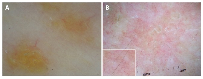
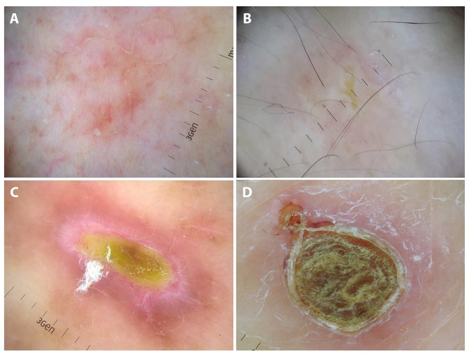
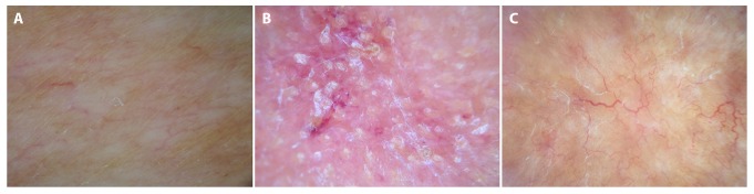
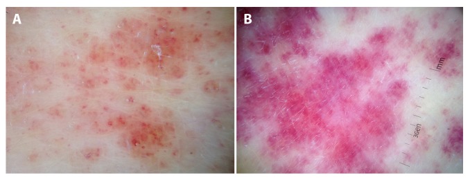
In addition to its use in pigmented and nonpigmented skin tumors, dermoscopy is gaining appreciation in assisting the diagnosis of nonneoplastic diseases, especially inflammatory dermatoses (inflammoscopy). In this field, dermoscopic examination should be considered as the second step of a "2-step procedure," always preceded by the establishment of a differential diagnosis on the basis of clinical examination. In this paper, we sought to provide an up-to-date overview on the use of dermoscopy in common inflammatory dermatoses based on the available literature data. For practical purposes, the analyzed dermatoses are grouped according to the clinical presentation pattern, in line with the 2-step procedure principle: erythematous-desquamative and papulosquamous dermatoses, papulokeratotic dermatoses, erythematous facial dermatoses, sclero-atrophic dermatoses, and miscellaneous.

Keywords: dermoscopy; differential diagnosis; general dermatology; inflammoscopy.


Conflict of interest statement

Competing interests: The author has no conflicts of interest to disclose.

Figures

Figure 1
Figure 1
Uniform dotted vessels and diffuse white scales in psoriasis (A). Dotted vessels distributed in clusters and yellow scales and serocrusts in eczematous dermatitis (B). The typical Wickham striae of lichen planus; brownish dots are also visible around Wickham striae (C). Lichen nitidus typically reveals roundish, well-defined, white areas devoid of physiological skin markings (D). [Copyright: ©2019 Errichetti.]
Figure 2
Figure 2
Dermoscopy of pityriasis rosea shows the characteristic peripheral whitish scaling (“collarette” sign). No vessels are seen (A); pityriasis rosea in an atopic patient displays yellow serocrusts/scaling along with the peripheral whitish scaling collarette (B). Central amorphous brownish crust surrounded by a peripheral scaling collarette and a purpuric halo is visible in a case of pityriasis lichenoides et varioliformis acuta (C). Dermoscopic examination of pityriasis lichenoides chronica reveals an orange structureless area along with nondotted vessels (linear-irregular and branching vessels), white scaling, and sparse dotted vessels (D). [Copyright: ©2019 Errichetti.]
Figure 3
Figure 3
Pityriasis rubra pilaris features a roundish, yellowish area surrounded by vessels of mixed morphology (ie, linear and dotted) (A). Dermoscopy in subacute lupus erythematosus reveals white scales and a mixed vascular pattern (ie, dotted and linear vessels in this case; visualized more clearly in the box) over a pinkish-reddish background (B). [Copyright: ©2019 Errichetti.]
Figure 4
Figure 4
The typical white keratotic track having 2 free edges is visible in a case of porokeratosis (A). Darier disease characteristically reveals a central star-like, yellowish area surrounded by a peripheral white halo (B). The “white starburst” pattern (peripheral radial white striae over a reddish-brownish background) is visible in a case of prurigo nodularis; a central yellow crust is also present (C). Acquired reactive perforating collagenosis displays a central round brownish-greenish structureless area surrounded by a white keratotic collarette and an erythematous halo (“trizonal concentric” pattern) (D). [Copyright: ©2019 Errichetti.]
Figure 5
Figure 5
Erythemato-telangiectatic rosacea with its typical linear vessels arranged in polygonal networks (vascular polygons) (A). The so-called Demodex tails (white-yellowish, protruding, follicular keratotic plugs) are visible in demodicosis (B). White keratotic plugs over a reddish background are the typical dermoscopic features of active discoid lupus erythematosus (C). Dermoscopy of sarcoidosis reveals orange structureless areas with overlying focused linear vessels; white areas are also visible (D). [Copyright: ©2019 Errichetti.]
Figure 6
Figure 6
Morphea is characterized by dull white areas with blurry margins (“white clouds”) (A). Keratotic follicular plugs, white scales, and hemorrhagic spots over a white-pinkish background are visible in lichen sclerosus (B). Necrobiosis lipoidica: yellow-orange structureless areas and branching-serpentine vessels whose diameter decreases from the center to the periphery of the lesion (C). [Copyright: ©2019 Errichetti.]
Figure 7
Figure 7
Schamberg disease: focused reddish purpuric dots and globules over a coppery background (A). Unlike capillaritis, dermoscopy of small-vessel skin vasculitis reveals blurrier violaceous purpuric globules (B). [Copyright: ©2019 Errichetti.]
Figure 8
Figure 8
Idiopathic guttate hypomelanosis: well-defined roundish homogeneous whitish area (“cloudy” pattern) surrounded by patchy hyperpigmented network (A). Vitiligo: white areas and perifollicular pigmentation (B). [Copyright: ©2019 Errichetti.]
Figure 9
Figure 9
Brownish dots in lichen pigmentosus (A). In ashy dermatosis dots are smaller and bluish (B). [Copyright: ©2019 Errichetti.]
Figure 10
Figure 10
Coarse brownish network in urticaria pigmentosa (A). Telangiectasia macularis eruptiva perstans reveals tortuous linear vessels on a brownish base (B). [Copyright: ©2019 Errichetti.]
Figure 11
Figure 11
In both “palisading granuloma” and “interstitial” histological variants of granuloma annulare, dermoscopy shows unfocused vessels having a variable morphology (dotted, linear-irregular, and/or branching) over a more or less evident pinkish-reddish background (A,B). However, the former variant also features yellowish orange areas (A). [Copyright: ©2019 Errichetti.]

References

    1. Errichetti E, Stinco G. Dermoscopy in general dermatology: a practical overview. Dermatol Ther (Heidelb) 2016;6(4):471–507. - PMC - PubMed
    1. Errichetti E, Stinco G. The practical usefulness of dermoscopy in general dermatology. G Ital Dermatol Venereol. 2015;150(5):533–546. - PubMed
    1. Errichetti E, Zalaudek I, Kittler H, et al. Standardization of dermoscopic terminology and basic dermoscopic parameters to evaluate in general dermatology (non-neoplastic dermatoses): an expert consensus on behalf of the International Dermoscopy Society. Br J Dermatol. 2019 May 11; doi: 10.1111/bjd.18125. Epub ahead of print. - DOI - PubMed
    1. Errichetti E, Stinco G. Clinical and dermoscopic response predictors in psoriatic patients undergoing narrowband ultraviolet B phototherapy: results from a prospective study. Int J Dermatol. 2018;57(6):681–686. - PubMed
    1. Lallas A, Kyrgidis A, Tzellos TG, et al. Accuracy of dermoscopic criteria for the diagnosis of psoriasis, dermatitis, lichen planus and pityriasis rosea. Br J Dermatol. 2012;166(6):1198–1205. - PubMed

LinkOut - more resources