. 2019 Jul;9(4):690-701.
doi: 10.1016/j.apsb.2019.03.003. Epub 2019 Mar 16.

Matrine attenuates oxidative stress and cardiomyocyte apoptosis in doxorubicin-induced cardiotoxicity via maintaining AMPK α/UCP2 pathway

Affiliations

Matrine attenuates oxidative stress and cardiomyocyte apoptosis in doxorubicin-induced cardiotoxicity via maintaining AMPK α/UCP2 pathway

Can Hu et al. Acta Pharm Sin B. 2019 Jul.

Abstract

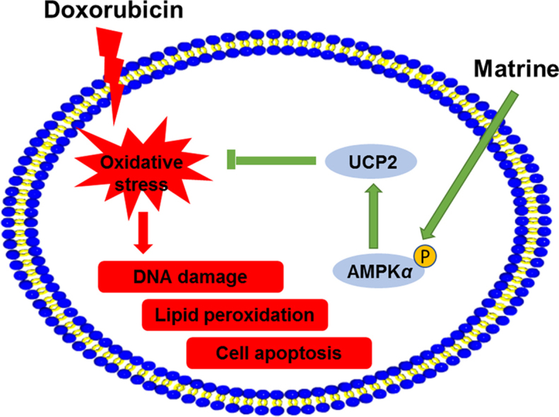
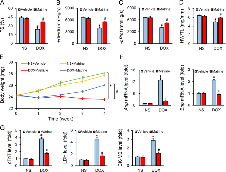
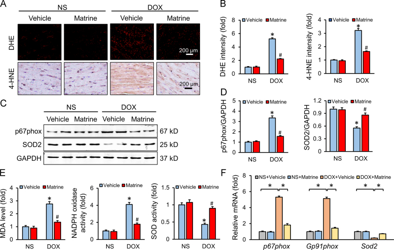
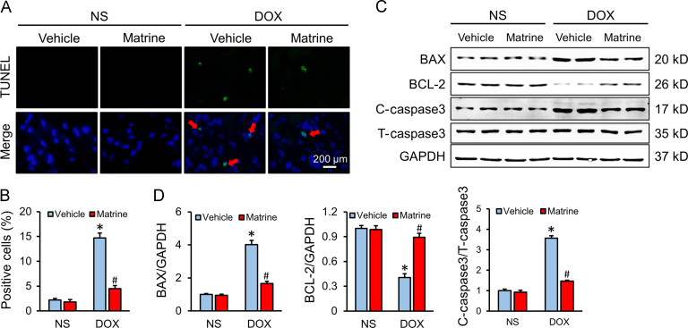
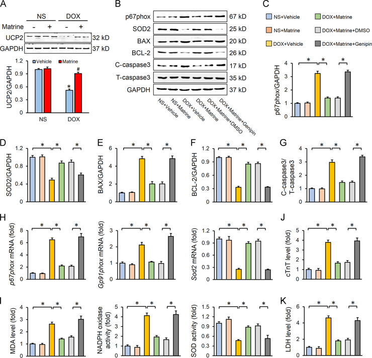
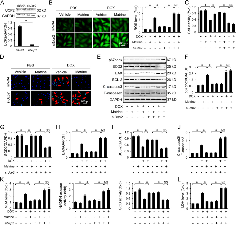
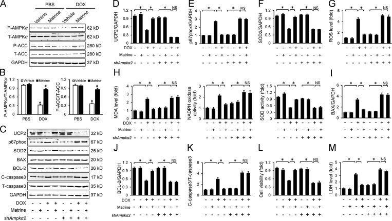
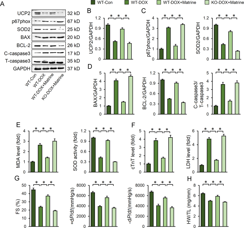
Oxidative stress and cardiomyocyte apoptosis are involved in the pathogenesis of doxorubicin (DOX)-induced cardiotoxicity. Matrine is well-known for its powerful anti-oxidant and anti-apoptotic capacities. Our present study aimed to investigate the effect of matrine on DOX-induced cardiotoxicity and try to unearth the underlying mechanisms. Mice were exposed with DOX to generate DOX-induced cardiotoxicity or normal saline as control. H9C2 cells were used to verify the effect of matrine in vitro. DOX injection triggered increased generation of reactive oxygen species (ROS) and excessive cardiomyocyte apoptosis, which were significantly mitigated by matrine. Mechanistically, we found that matrine ameliorated DOX-induced uncoupling protein 2 (UCP2) downregulation, and UCP2 inhibition by genipin could blunt the protective effect of matrine on DOX-induced oxidative stress and cardiomyocyte apoptosis. Besides, 5'-AMP-activated protein kinase α2 (Ampkα2) deficiency inhibited matrine-mediated UCP2 preservation and abolished the beneficial effect of matrine in mice. Besides, we observed that matrine incubation alleviated DOX-induced H9C2 cells apoptosis and oxidative stress level via activating AMPKα/UCP2, which were blunted by either AMPKα or UCP2 inhibition with genetic or pharmacological methods. Matrine attenuated oxidative stress and cardiomyocyte apoptosis in DOX-induced cardiotoxicity via maintaining AMPKα/UCP2 pathway, and it might be a promising therapeutic agent for the treatment of DOX-induced cardiotoxicity.

Keywords: 4-HNE, 4-hydroxynonenal; ACC, acetyl-CoA carboxylase; AMPKα; AMPKα, 5′-AMP-activated protein kinase α; ANOVA, analysis of variance; Apoptosis; BAX, BCL-2-associated X protein; BCA, bicinchoninic acid; BCL-2, B-cell lymphoma 2; C-caspase 3, cleaved-caspase3; CCK-8, cell counting kit 8; CK-MB, creatine kinase isoenzymes; DCFH-DA, 2′,7′-dichlorodihydrofluorescein diacetate; DHE, dihydroethidium; DMEM, Dulbecco׳s modified Eagle׳s medium; DOX, doxorubicin; FBS, fetal bovine serum; FS, fractional shortening; GAPDH, glyceraldehyde 3-phosphate dehydrogenase; HW, heart weight; LDH, lactate dehydrogenase; MDA, malondialdehyde; Matrine; Oxidative stress; PPARs, peroxisomal proliferators-activated receptors; ROS, reactive oxygen species; SOD2, superoxide dismutase 2; T-caspase3, total-caspase3; TL, tibia length; TUNEL, TdT-mediated dUTP nick end-labelling; Top2, topoisomerase-II; UCP2; UCP2, uncoupling protein 2; cTnT, cardiac isoform of Tropnin T.


Figures

fx1
Graphical abstract
Fig. 1
Figure 1
Matrine attenuated doxorubicin (DOX)-induced cardiotoxicity in mice. (A) Quantitative analysis of fraction shortening (FS) obtained from echocardiography (n = 8). (B) and (C) Hemodynamic analysis of mice with or without matrine treatment (n = 8). (D) Statistical results of the heart weight (HW)/tibia length (TL) (n = 12). (E) Body weight of four groups (n = 12). (F) Relative mRNA levels of Anp, Bnp (n = 6). (G) Plasma levels of cTnT, LDH and CK-MB in mice (n = 6). Values represent mean ± SEM. *P< 0.05 versus NS+Vehicle; #P< 0.05 versus DOX+Vehicle.
Fig. 2
Figure 2
Matrine prevented oxidative damage in response to DOX insult. (A) and (B) Representative DHE and 4-HNE staining images and the statistical results (n = 6). (C) and (D) Representative Western blot and quantitative data (n = 6). (E) The content of MDA, NADPH oxidase activity and total SOD activity in the myocardium (n = 6). (F) Relative mRNA levels of p67phox, Gp91phox and Sod2 (n = 6). Values represent the mean±SEM. *P< 0.05 versus NS+Vehicle; #P< 0.05 versus DOX+Vehicle.
Fig. 3
Figure 3
Matrine attenuated DOX-induced cardiomyocyte apoptosis. (A) and (B) Representative TUNEL staining images and the quantitative results for apoptosis in heart tissues (n = 6). (C) and (D) Western blot and statistical results in the indicated groups (n = 6). Red arrows indicate TUNEL positive staining. Values represent the mean±SEM. *P< 0.05 versus NS+Vehicle; #P< 0.05 versus DOX+Vehicle.
Fig. 4
Figure 4
UCP2 was responsible for matrine-mediated beneficial effect on DOX-induced cardiotoxicity. (A) UCP2 expression level in murine hearts with or without DOX or matrine (n = 6; *P< 0.05 versus NS+Vehicle, #P< 0.05 versus DOX+Vehicle). (B)–(G) Western blot and statistical results in the indicated groups (n = 6). (H) Relative mRNA levels of p67phox, Gp91phox and Sod2 (n = 6). (I) The content of MDA, NADPH oxidase activity and total SOD activity in the myocardium (n = 6). (J)–(K) Plasma levels of cTnT and LDH (n = 6). Values represent the mean±SEM. *P< 0.05 versus the matched groups.
Fig. 5
Figure 5
Knockdown of UCP2 blunted the protective effects of matrine in vitro. (A) Western blot for UCP2 and the quantitative data for H9C2 cells (n = 6). (B) Representative DCFH-DA images and the statistical results. (C) CCK-8 assay for cell viability (n = 5). (D) Representative images of TUNEL in H9C2 cells (n = 5). (E)–(J) Western blot and statistical results (n = 6). (K) The content of MDA, NADPH oxidase activity and total SOD activity in H9C2 cells (n = 5). (L) LDH levels in H9C2 cells (n = 5). Red arrows indicate TUNEL positive staining. Values represent the mean±SEM. *P< 0.05 versus the matched groups. NS indicates no statistical difference.
Fig. 6
Figure 6
Matrine prevented DOX-induced downregulation of UCP2 via activating AMPKα. (A) and (B) Western blot and statistical results (n = 6). (C)–(F) Western blot and statistical results (n = 6). (G) Statistical results of intracellular ROS production detected by DCFH-DA in H9C2 cells (n = 5). (H) The content of MDA, NADPH oxidase activity and total SOD activity in H9C2 cells (n = 5). (I)–(K) Western blot and statistical results (n = 6). (L) Cell viability by CCK-8 (n = 5). (M) Relative LDH levels (n = 5). Values represent the mean±SEM. *P< 0.05 versus the matched groups. NS indicates no statistical difference.
Fig. 7
Figure 7
Ampkα deficiency abolished the protective effect of matrine in vivo. (A)–(D) Representative Western blot images and statistical results (n = 6). (E) The content of MDA and total SOD activity (n = 6). (F) Plasma levels cTnT and LDH (n = 6). (G) Heart function evaluated by echocardiography and hemodynamic analysis of mice (n = 8). (H) Statistical results of the HW/TL (n = 12). Values represent the mean±SEM. *P< 0.05 versus the matched groups.

References

    1. Carvalho C., Santos R.X., Cardoso S., Correia S., Oliveira P.J., Santos M.S. Doxorubicin: the good, the bad and the ugly effect. Curr Med Chem. 2009;16:3267–3285. - PubMed
    1. Li M., Sala V., De Santis M.C., Cimino J., Cappello P., Pianca N. Phosphoinositide 3-Kinase γ inhibition protects from anthracycline cardiotoxicity and reduces tumor growth. Circulation. 2018;138:696–711. - PubMed
    1. Yamanaka S., Tatsumi T., Shiraishi J., Mano A., Keira N., Matoba S. Amlodipine inhibits doxorubicin-induced apoptosis in neonatal rat cardiac myocytes. J Am Coll Cardiol. 2003;41:870–878. - PubMed
    1. Pacher P., Liaudet L., Bai P., Mabley J.G., Kaminski P.M., Virag L. Potent metalloporphyrin peroxynitrite decomposition catalyst protects against the development of doxorubicin-induced cardiac dysfunction. Circulation. 2003;107:896–904. - PubMed
    1. Wang W., Fang H., Groom L., Cheng A., Zhang W., Liu J. Superoxide flashes in single mitochondria. Cell. 2008;134:279–290. - PMC - PubMed