. 2019 Jul;9(4):745-757.
doi: 10.1016/j.apsb.2019.02.006. Epub 2019 Feb 22.

Silibinin ameliorates hepatic lipid accumulation and oxidative stress in mice with non-alcoholic steatohepatitis by regulating CFLAR-JNK pathway

Affiliations

Silibinin ameliorates hepatic lipid accumulation and oxidative stress in mice with non-alcoholic steatohepatitis by regulating CFLAR-JNK pathway

Yayun Liu et al. Acta Pharm Sin B. 2019 Jul.

Abstract

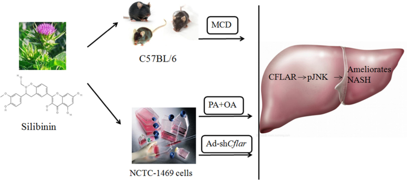
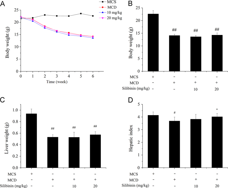
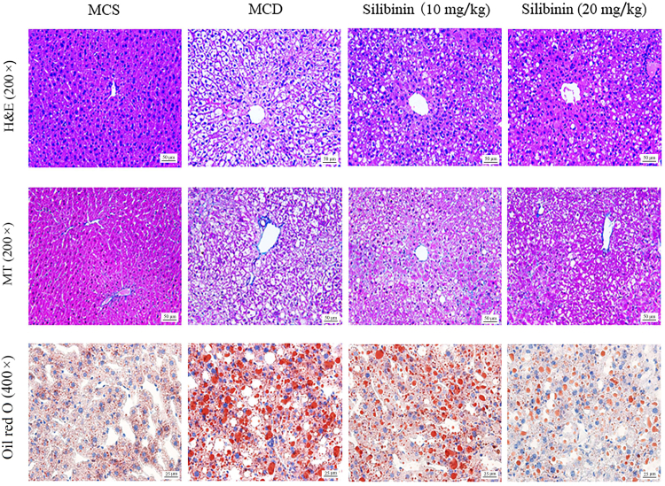
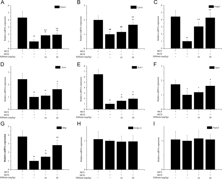
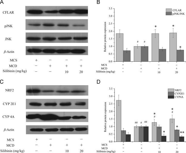
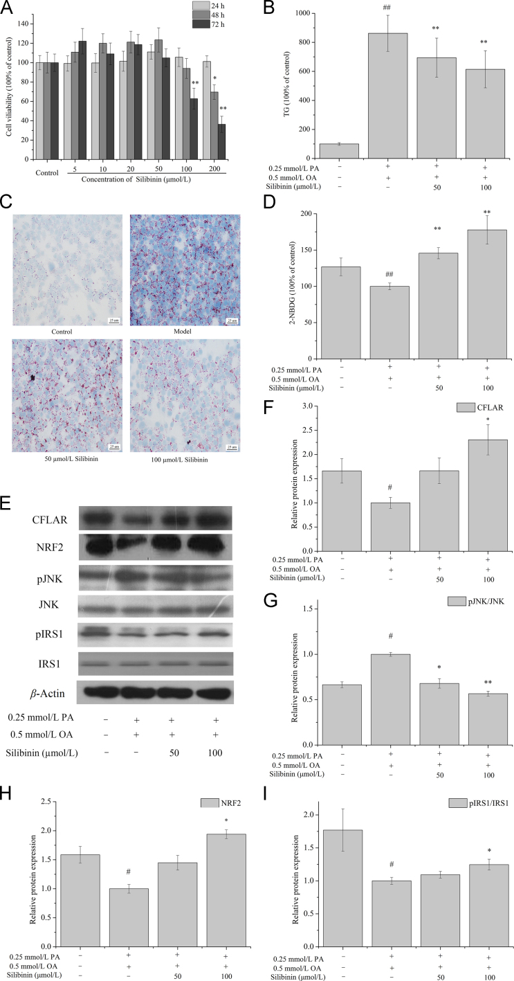
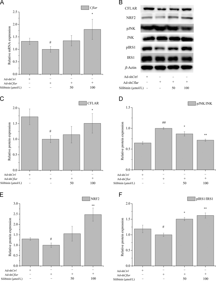
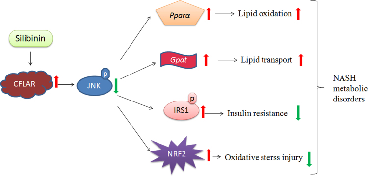
Non-alcoholic steatohepatitis (NASH) is a chronic metabolic syndrome and the CFLAR-JNK pathway can reverse the process of NASH. Although silibinin is used for the treatment of NASH in clinical, its effect on CFLAR-JNK pathway in NASH remains unclear. This study aimed to investigate the effect of silibinin on CFLAR-JNK pathway in NASH models both in vivo and in vitro. The in vivo study was performed using male C57BL/6 mice fed with methionine- choline-deficient diet and simultaneously treated with silibinin for 6 weeks. The in vitro study was performed by using mouse NCTC-1469 cells which were respectively pretreated with oleic acid plus palmitic acid, and adenovirus-down Cflar for 24 h, then treated with silibinin for 24 h. After the drug treatment, the key indicators involved in CFLAR-JNK pathway including hepatic injury, lipid metabolism and oxidative stress were determined. Silibinin significantly activated CFLAR and inhibited the phosphorylation of JNK, up-regulated the mRNA expression of Pparα, Fabp5, Cpt1α, Acox, Scd-1, Gpat and Mttp, reduced the activities of serum ALT and AST and the contents of hepatic TG, TC and MDA, increased the expression of NRF2 and the activities of CAT, GSH-Px and HO-1, and decreased the activities and expression of CYP2E1 and CYP4A in vivo. These effects were confirmed by the in vitro experiments. Silibinin prevented NASH by regulating CFLAR-JNK pathway, and thereby on one hand promoting the β-oxidation and efflux of fatty acids in liver to relieve lipid accumulation, and on the other hand inducing antioxidase activity (CAT, GSH-Px and HO-1) and inhibiting pro-oxidase activity (CYP2E1 and CYP4A) to relieve oxidative stress.

Keywords: 2-NBDG, 2-(N-(7-nitrobenz-2-oxa-1,3-diazol-4-yl) amino)-2-deoxyglucose; ALT, alanine aminotransferase; AST, aspartate aminotransferase; Acox, acyl-coenzyme A oxidase X; Akt, serine–threonine protein kinase; CAT, catalase; CFLAR; CFLAR, caspase 8 and Fas-associated protein with death domain-like apoptosis regulator; CYP2E1, cytochrome P450 2E1; CYP4A, cytochrome P450 4A; Cpt1α, carnitine palmitoyl transferase 1α; Fabp5, fatty acid-binding proteins 5; GSH-Px, glutathione peroxidase; Gpat, glycerol-3-phosphate acyltransferase; HE, hematoxylin–eosin; HO-1, heme oxygenase 1; IR, insulin resistance; IRS1, insulin receptor substrate 1; JNK, c-Jun N-terminal kinase; Lipid accumulation; MAPK, mitogen-activated protein kinase; MCD, methionine- and choline-deficient; MCS, methionine- and choline-sufficient; MDA, malondialdehyde; MT, Masson–Trichrome; Mttp, microsomal triglyceride transfer protein; NAFLD, non-alcoholic fatty liver disease; NASH; NASH, nonalcoholic steatohepatitis; NF-κB, nuclear factor κB; NRF2, nuclear factor erythroid 2-related factor 2; OA, oleic acid; ORO, oil red O; Oxidation stress; PA, palmitic acid; PI3K, phosphatidylinositol 3-hydroxy kinase; Pnpla3, phospholipase domain containing 3; Pparα, peroxisome proliferator activated receptor α; SD, Sprague–Dawley; Scd-1, stearoyl-coenzyme A desaturase-1; Silibinin; Srebp-1c, sterol regulatory element binding protein-1C; TC, total cholesterol; TG, triglyceride; pIRS1, phosphorylation of insulin receptor substrate 1; pJNK, phosphorylation of c-Jun N-terminal kinase.


Figures

fx1
Graphical abstract
Fig. 1
Figure 1
Effects of silibinin on body weight, liver weight and hepatic index in different groups of mice. C57BL/6 were treated with MCD and silibinin for 6 weeks and the body weight during 6 weeks (A), the body weight (B), liver weight (C) and hepatic index (D) after 6 weeks in different groups of mice were detected. Values (mean ± SD) were obtained from each group (n = 8) after 6 weeks of the experimental period. #P < 0.05 and ##P < 0.01 versus MCS group; *P < 0.05 and **P < 0.01 versus MCD group. MCD, methionine- and choline-deficient; MCS, methionine- and choline-sufficient.
Fig. 2
Figure 2
Effects of silibinin on hepatic histological changes. H&E, MT and oil red O staining of liver sections in different groups of mice. HE, hematoxylin and eosin MCD, methionine- and choline-deficient; MCS, methionine- and choline-sufficient; MT, Masson–Trichrome.
Fig. 3
Figure 3
Effects of silibinin on the mRNA expression of hepatic lipid accumulation-related genes in different groups of mice. Values (mean ± SD) were obtained from each group (n = 8) after 6 weeks of the experimental period. #P < 0.05 and ##P < 0.01 versus MCS group; *P < 0.05 and **P < 0.01 versus MCD group. Acox, Acyl-coenzyme A oxidase X; Cflar, caspase 8 and Fas-associated protein with death domain-like apoptosis regulator; Cpt1α, carnitine palmitoyl transferase 1α; Fabp5, fatty acid-binding proteins 5; Gpat, glycerol-3-phosphate acyltransferase; MCD, methionine- and choline-deficient; MCS, methionine- and choline-sufficient; Mttp, microsomal triglyceride transfer protein; Pnpla3, phospholipase domain containing 3; Pparα, peroxisome proliferator activated receptor α; Scd-1, stearoyl-coenzyme A desaturase-1; Srebp-1c, sterol regulatory element binding protein-1C.
Fig. 4
Figure 4
Effects of silibinin on the protein expression of hepatic CFLAR-JNK pathway related genes in different groups of mice. Values (mean ± SD) were obtained from each group (n = 8) after 6 weeks of the experimental period. #P < 0.05 and ##P < 0.01 versus MCS group; *P < 0.05 and **P < 0.01 versus MCD group. CFLAR, caspase 8 and Fas-associated protein with death domain-like apoptosis regulator; JNK, c-Jun N-terminal kinase; MCD, methionine- and choline-deficient; MCS, methionine- and choline-sufficient; NRF2, nuclear factor erythroid 2-related factor 2; pJNK, phosphorylation of c-Jun N-terminal kinase.
Fig. 5
Figure 5
Effects of silibinin on cellular TG and 2-NBDG contents, and the protein expression of CFLAR, pJNK/JNK, NRF2 and pIRS1/IRS1 in OA and PA pretreated NCTC-1469 cells. NCTC-1469 cells were pretreated with OA and PA and then treated with silibinin to detected the cellular viability (A), TG (B), oil red O staining (C), 2-NBDG (D), protein expression of CFLAR, NRF2, pJNK/JNK and pIRS1/IRS1 (E)–(I). The representative results from three independent experiments were shown. Cellular protein expression levels were analyzed by Western blot. The relative protein levels were determined after normalization with β-actin. The data were expressed as the means ± SD. #P < 0.05 and ##P < 0.01 versus control group; *P < 0.05 and **P < 0.01 versus model group. Scale bar=25 μm. CFLAR, caspase 8 and Fas-associated protein with death domain-like apoptosis regulator; IRS1, insulin receptor substrate 1; JNK, c-Jun N-terminal kinase; NRF2, nuclear factor erythroid 2-related factor 2; OA, oleic acid; PA, palmitic acid; pIRS1, phosphorylation of insulin receptor substrate 1; pJNK, phosphorylation of c-Jun N-terminal kinase; TG, triglyceride; 2-NBDG, 2-(N-(7-nitrobenz-2-oxa-1,3-diazol-4-yl) amino)-2-deoxyglucose.
Fig. 6
Figure 6
Effects of silibinin on the expression of CFLAR-JNK pathway-related genes in NCTC-1469 cells after adenovirus-mediated knock-down of Cflar. NCTC-1469 cells were transfected with adenovirus and then treated with silibinin to detected the cellular mRNA expression of Cflar (A), protein expression of CFLAR, pJNK/JNK NRF2, and pIRS1/IRS1 (B)–(F). The representative results from three independent experiments were shown. Cellular protein expression levels were analyzed by Western blot. The relative protein levels were determined after normalization with β-actin. The data were expressed as the means ± SD. #P < 0.05 and ##P < 0.01 versus control group; *P < 0.05 and **P < 0.01 versus model group. CFLAR, caspase 8 and Fas-associated protein with death domain-like apoptosis regulator; IRS1, insulin receptor substrate 1; JNK, c-Jun N-terminal kinase; NRF2, nuclear factor erythroid 2-related factor 2; pIRS1, phosphorylation of insulin receptor substrate 1; pJNK, phosphorylation of c-Jun N-terminal kinase.
Fig. 7
Figure 7
Proposed mechanism of silibinin against NASH. NASH, nonalcoholic steatohepatitis.

References

    1. Kleiner D.E., Brunt E.M., van Natta M., Behling C., Contos M.J., Cummings O.W. Design and validation of a histoloical scoring system for nonalcoholic fatty liver disease. Hepatology. 2005;411:313–321. - PubMed
    1. Michelotti G.A., Machado M.V., Diehl A.M. NAFLD, NASH and liver cancer. Nat Rev Gastroenterol Hepatol. 2013;10:656–665. - PubMed
    1. Byrne C.D., Targher G. NAFLD: a multisystem disease. J Hepatol. 2015;62:S47–S64. - PubMed
    1. Zhu Y., Liu H., Zhang M., Guo G.L. Fatty liver diseases, bile acids, and FXR. Acta Pharm Sin B. 2016;6:409–412. - PMC - PubMed
    1. Nascimbeni F., Pais R., Bellentani S., Day C.P., Ratziu V., Loria P. From NAFLD in clinical practice to answers from guidelines. J Hepatol. 2013;59:859–871. - PubMed

LinkOut - more resources