. 2019 Jul;9(4):819-831.
doi: 10.1016/j.apsb.2019.01.018. Epub 2019 Jan 31.

Multifunctional polymeric micelle-based chemo-immunotherapy with immune checkpoint blockade for efficient treatment of orthotopic and metastatic breast cancer

Affiliations

Multifunctional polymeric micelle-based chemo-immunotherapy with immune checkpoint blockade for efficient treatment of orthotopic and metastatic breast cancer

Jiaojie Wei et al. Acta Pharm Sin B. 2019 Jul.

Abstract

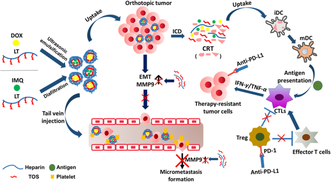
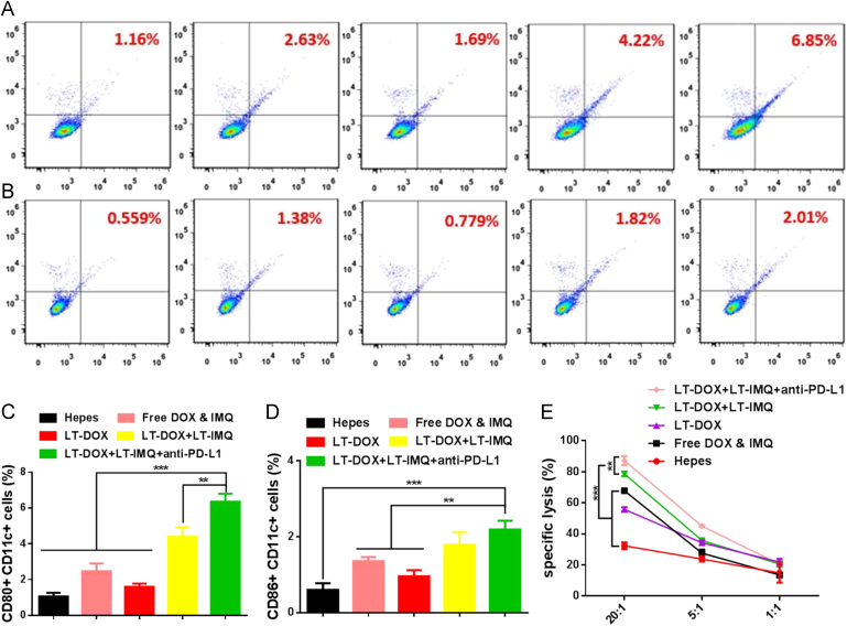
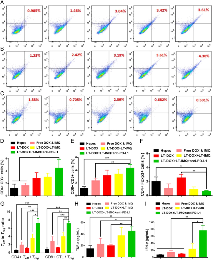
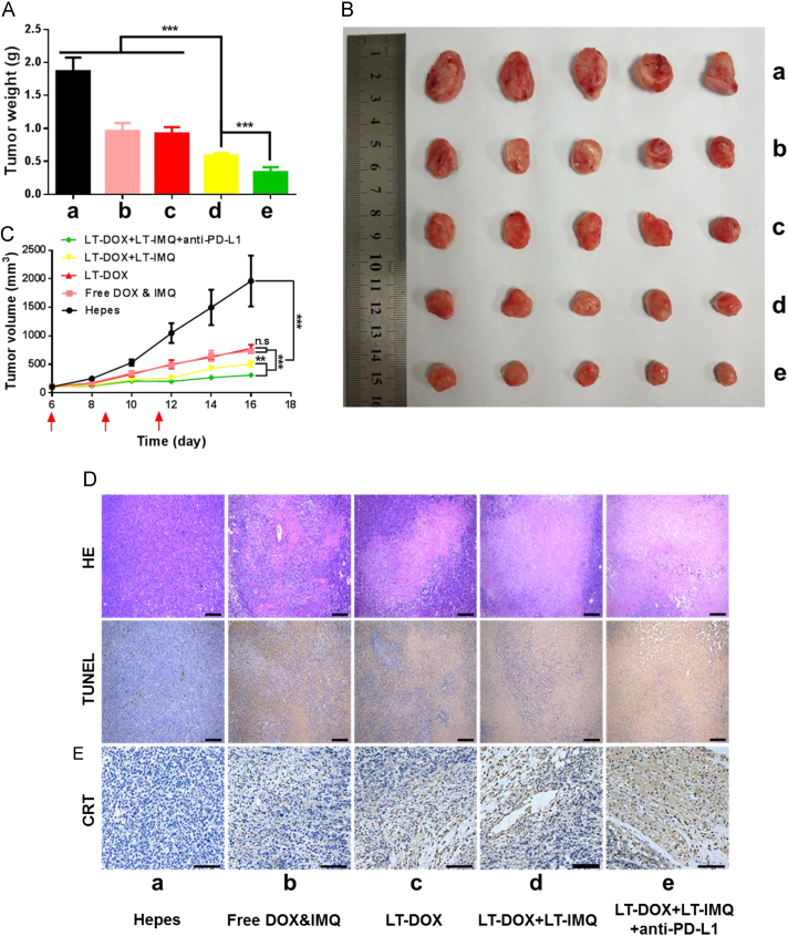
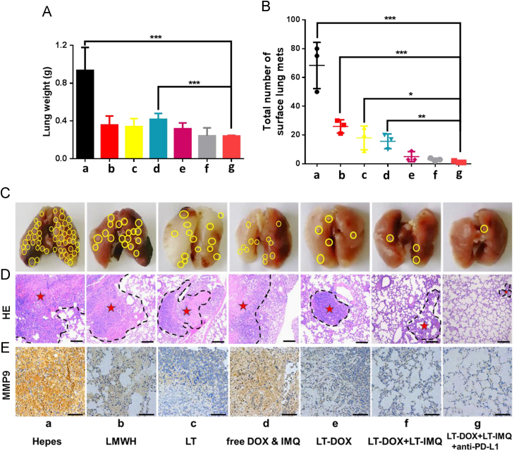
Immunotherapy has become a highly promising paradigm for cancer treatment. Herein, a chemo-immunotherapy was developed by encapsulating chemotherapeutic drug doxorubicin (DOX) and Toll-like receptor 7 agonist imiquimod (IMQ) in low molecular weight heparin (LMWH)-d-α-tocopheryl succinate (TOS) micelles (LT). In this process, LMWH and TOS were conjugated by ester bond and they were not only served as the hydrophilic and hydrophobic segments of the carrier, but also exhibited strong anti-metastasis effect. The direct killing of tumor cells mediated by DOX-loaded micelles (LT-DOX) generated tumor-associated antigens, initiating tumor-specific immune responses in combination with IMQ-loaded micelles (LT-IMQ). Furthermore, the blockade of immune checkpoint with programmed cell death ligand 1 (PD-L1) antibody further elevated the immune responses by up-regulating the maturation of DCs as well as the ratios of CD8+ CTLs/Treg and CD4+ Teff/Treg. Therefore, such a multifunctional strategy exhibited great potential for inhibiting the growth of orthotopic and metastatic breast cancer.

Keywords: Anti-metastasis; Checkpoint blockade; Immunogenic cell death; Immunotherapy; Nanoparticle.


Figures

fx1
Graphical abstract
Fig. 1
Figure 1
Hydrodynamic diameters and TEM image of (A) LT-DOX and (B) LT-IMQ. (C) Photo images of plastic pipe containing supernatant from red blood cell content treated with different concentrations of LT. (D) Hemolysis (%) of red blood cell cultured with LT at various concentrations. Less than 5% hemolysis was regarded as nontoxic. (E) Stability of nanoparticles represented by size when incubated in 50% FBS for 48 h at 37 °C. (F) The turbidity variation (represented by transmittance) of nanoparticles in 50% FBS. (G) In vitro release kinetics of DOX and IMQ from micelles of LT-DOX and LT-IMQ.
Fig. 2
Figure 2
(A) Representative images of healing extent treated with different preparations after 24 h. (B) Wound healing rate. (C) Representative images of invaded 4T1 cells when treated with different formulations for 48 h. (D) Relative invasion rate of invaded cells. Data are mean ± SD, n = 3; *P < 0.05; **P < 0.01; and ***P < 0.001.
Fig. 3
Figure 3
(A) CRT exposure on the cell surface of 4T1 cells was evaluated by flow cytometry after different treatments. (B) Immunofluorescence microscopy of CRT expression on the cell surface of 4T1 cells after different treatments. The scale bar represents 10 μm. (C) and (D) DOX and IMQ plasma concentration–time profiles in BALB/c mice treated with different DOX and IMQ formulations (at a dose of 3 mg/kg DOX, 0.75 mg/kg IMQ; data are mean ± SD, n = 3).
Fig. 4
Figure 4
The expression status of CD80+ and CD86+ on CD11c+ dendritic cells from spleens. Representative flow cytometry plots showing percentages of CD11c+CD80+ cells (A) and CD11c+CD86+ cells (B). From left to right are groups of Hepes, free DOX & IMQ, LT-DOX, LT-DOX+LT-IMQ, LT-DOX+LT-IMQ+anti-PD-L1. Proportions of CD11c+CD80+ cells (C) and CD11c+CD86+ cells (D). (E) The cytotoxicity assay of CD8+ T lymphocytes against 4T1 cells. Data are mean ± SD, n = 3; *P < 0.05; **P < 0.01; and ***P < 0.001.
Fig. 5
Figure 5
Representative flow cytometry plots showing percentages of tumor-infiltrating CD3e+CD4+ killer T cells (A), CD3e+CD8+ effector T cells (B) and CD4+FoxP3+ regulatory T cells (C) in solid tumors after various treatments indicated. From left to right are groups of Hepes, free DOX & IMQ, LT-DOX, LT-DOX+LT-IMQ, LT-DOX+LT-IMQ+anti-PD-L1. Proportions of CD3e+CD4+ effector T cells (C), CD3e+CD8+ T cells (D) and CD4+FoxP3+ regulatory T cells (E). (G) CD4+CTLs/Treg ratios and CD8+ effector T cells/Treg cells ratios in the solid tumors upon various treatments. Cytokine levels of TNF-α (H), and IFN-γ (I) in sera from mice isolated on day 3 after various treatments. Three mice were measured in each group in A–I. Data are mean ± SD, n = 3; *P < 0.05; **P < 0.01; and ***P < 0.001.
Fig. 6
Figure 6
(A) The weights of dissected tumors at the end of treatment. (B) Tumor growth curves of 4T1 tumors when treated with different formulations. (C) Representative picture of tumors 16 days after implantation of 4T1 cells. (D) Representative H&E and TUNEL staining of tumor sections. The scale bar represents 200 μm. (E) Immunohistochemistry to detect CRT in tumors (shown in brown). The scale bar represents 100 μm. Data are mean ± SD, n = 6; *P < 0.05; **P < 0.01; and ***P < 0.001.
Fig. 7
Figure 7
(A) The weights of lungs at the end of treatment. (B) Total number of surface lung Mets. (C) Representative pictures of lungs 23 days after injection of 4T1 cells via tail vein. The yellow circle represents the Mets. (D) Histological analysis of lung sections. The scale bar represents 200 μm. The dashed lines mean metastasis loci. (E) Immunohistochemistry to detect MMP9 in lung (shown in brown). The scale bar represents 100 μm. Data are mean ± SD, n = 5; *P < 0.05; **P < 0.01; and ***P < 0.001.

References

    1. Seigel R., Naishadham D., Jemal A. Cancer statistics, 2013. CA Cancer J Clin. 2013;63:11–30. - PubMed
    1. Minotti G., Menna P., Salvatorelli E., Cairo G., Gianni L. Anthracyclines: molecular advances and pharmacologic developments in antitumor activity and cardiotoxicity. Pharmacol Rev. 2004;56:185–229. - PubMed
    1. Shen N., Hu J., Zhang L., Zhang L., Sun Y., Xie Y. Doxorubicin-loaded zein in situ gel for interstitial chemotherapy of colorectal cancer. Acta Pharm Sin B. 2012;2:610–614. - PubMed
    1. Cook A.M., Lesterhuis W.J., Nowak A.K., Lake R.A. Chemotherapy and immunotherapy: mapping the road ahead. Curr Opin Immunol. 2015;39:23–39. - PubMed
    1. Noelia C., Pequignot M.O., Antoine T., François G., Stéphan R., Nathalie C. Caspase-dependent immunogenicity of doxorubicin-induced tumor cell death. J Exp Med. 2005;202:1691–1701. - PMC - PubMed