Review
. 2019 Jul 31;9(3):e28.
doi: 10.5415/apallergy.2019.9.e28. eCollection 2019 Jul.

Signals from the various immune cells in promoting food allergy-induced eosinophilic esophagitis like disease

Affiliations
Review

Signals from the various immune cells in promoting food allergy-induced eosinophilic esophagitis like disease

Priscilia Lianto et al. Asia Pac Allergy. .

Abstract

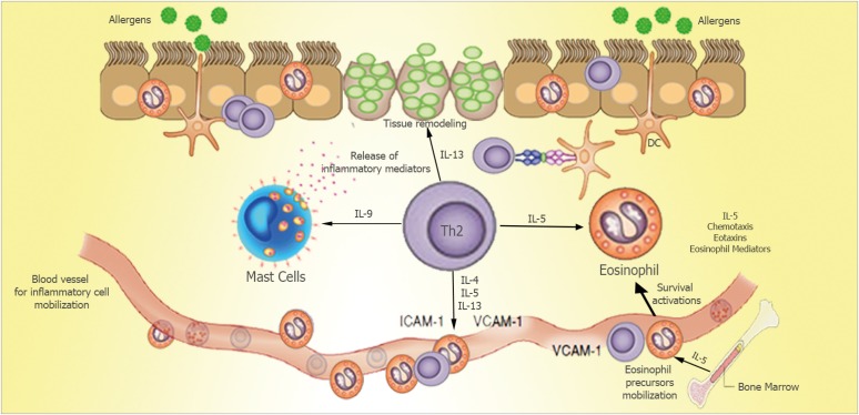
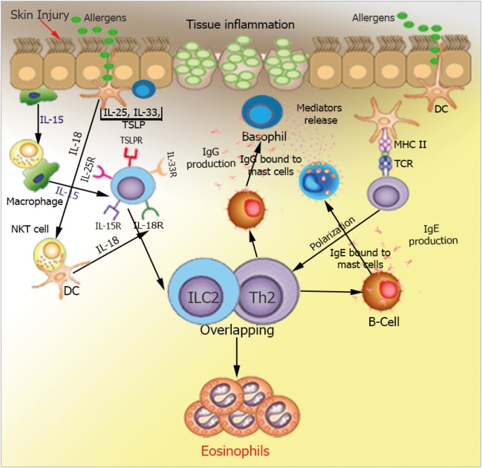
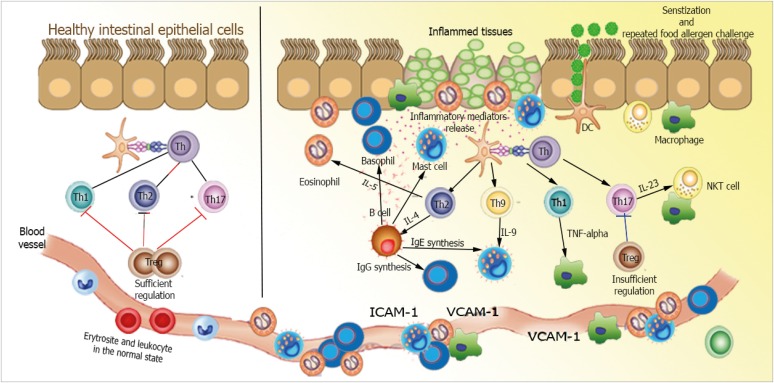
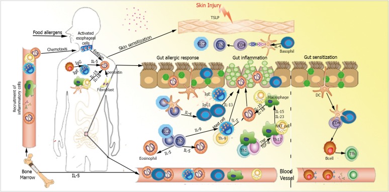
Eosinophilic esophagitis (EoE) is a recently recognized esophageal inflammatory disease with clinical manifestations arising from esophageal dysfunction. The etiology of EoE is currently being clarified and food allergy is evolving as the central cornerstone of EoE disease pathogenesis. Given the large number of eosinophils in the esophagus of people with EoE verified by data from murine models EoE is widely considered as the hallmark T-helper type 2 (Th2) disease of the esophagus. It is also known that some eosinophilic inflammation is controlled by other subsets of T cells such as Th9 or Th17 and control is also exerted by type 2 innate lymphoid cells acting together with basophils. In this paper we review results from molecular studies of mouse models in light of the results from the first clinical trials targeting key cytokines in humans and present in-depth molecular understanding of EoE.

Keywords: Elimination diet; Eosinophilic esophagitis; Food allergy; Pathogenesis; Therapy.


Figures

Fig. 1
Fig. 1. EoE exhibits features of a T-helper type 2 (Th2)-predominant inflammation. Antigen is captured by antigen presenting cells, which induces a Th2-response in eosinophilic esophagitis. Interleukin (IL)-5 stimulates the proliferation and differentiation of eosinophils in the bone marrow, retains their survival and enables their migration into the blood. IL-13 facilitates eotaxin-3 generation from the epithelial cells, which trafficking eosinophils to the esophagus. Eosinophils generate Th2-cytokines circulating the inflammatory cycle and, thus, release cytotoxic granules initiating tissue injury. They induce dendritic cells activation via eosinophil derived neurotoxin to trigger Th2-cells response. Eosinophils via major basic protein prime mast cells activation. Mast cells can also be actuated by antigens cross-linking their surface IgE. Activated mast cells release IL-13, IL-5 and other pro-inflammatory mediators. Together, mast cells and eosinophils cause tissue remodeling and inflammation. ICAM, intercellular adhesion molecule; VCAM, vascular cell adhesion molecule.
Fig. 2
Fig. 2. Comparative roles of T-helper type 2 (Th2) cells and type 2 innate lymphoid cells (ILC2) cells in 2 forms of eosinophilic esophagitis (EoE). Eosinophilic inflammation and tissue remodeling are compelled by adaptive Th2 cells that are stimulated by dendritic cells (DCs) to generate interleukin (IL)-5, IL-13 and IL-4, the latter driving IgE and IgG synthesis. ILC2 reacts to epithelial cytokines thymic stromal lymphopoietin (TSLP), IL-33, and IL-25 and in thusly deliver Th2 cytokines. ILC2 IL-4 play a role in contributing to Th2 cell differentiation. Additional ILC2 incitement may take place from prostaglandin D2 and leukotriene D4 mediators produced by mast cells and macrophages, and inhibitory signals from lipoxin A4. Th2 and ILC2 IL-5 generation promotes eosinophil activation and survival, whereas Th2 and ILC2 IL-13 leads to tissue remodeling, and along with IL-9, induces mucus production. Together, the responses triggered by secretion of type 2 cytokines from both ILC2 and Th2 cells orchestrate EoE allergic inflammation and tissue remodeling. NKT, natural killer T; MHC, major histocompatibility complex; TCR, T-cell receptor.
Fig. 3
Fig. 3. Abnormal interactions between allergen and the host immune system in gastrointestinal tissue in eosinophilic esophagitis (EoE) pathogenesis. Antigen presenting cells present food allergens as major histocompatibility complex class II conjugates to T-cell receptors on naive T cells. T cells then differentiate into T-helper type 1 (Th1) or Th17 cells, which secrete different pro-inflammatory and anti-inflammatory cytokines leading to recruitment of humoral and cellular factors of innate immunity. In healthy tissues (left), immune homeostasis is maintained by interdependent control exerted by sufficient Treg cells. In mucosal response in EoE (right), dysfunctional regulation of Th1 or Th17 pathways triggers an unregulated inflammatory response and recruitment of innate immune cells. Cytokine levels elevation can promote oxidative tissue damage and facilitate proteolytic peptides and enzymes recruitments, eventually presenting as gastrointestinal disease. Furthermore, enactment Th2 pathway can prompt plasma cells identifying fungal cell wall antigens and delivering antiglycan antibodies production. IL, interleukin; NKT, natural killer T; ICAM, intercellular adhesion molecule; VCAM, vascular cell adhesion molecule.
Fig. 4
Fig. 4. Food allergen as a promoter in eosinophilic esophagitis (EoE) pathogenesis. Notwithstanding with the history of genetic disposition or gastrointestinal disease, exposure to oral food allergen would promote the epithelial barrier disruption and affect the healthy balance of body microbiota in EoE subjects. Food allergens could then infiltrate also through the skin, engage to pathogen-related receptors and drive epithelial cells activation to pro-inflammatory cytokines generation which are responsible for the trafficking of the inflammatory cells. Under epithelial injury condition, thymic stromal lymphopoietin (TSLP) generation is provoked from epithelial cells. TSLP stimulates basophils to generate interleukin (IL)-4 which support allergic sensitization after allergen being presented to a naive T cell, driving allergen-specific Th2 cells activation. Adequate allergen challenges facilitate Th2 cells recruitment and expansion, secreting vast IL-5 and IL-13 which are crucial in eosinophils trafficking and tissue remodeling. Th2 cells locally promote class switching of B cells to produce antigen-specific IgE, which binds to the surface of mast cells and produce antigen-specific IgG which engage to the surface of basophils. Activation of mast cells and basophils leads to the release of pro-inflammatory mediators such as transforming growth factor beta (TGF-β), which contributes to the local inflammatory responses and promotes remodeling and enhances muscle cell contractility. Antigen presenting cell capturing food allergens would subsequently migrate to the regional lymph nodes and thus mediating food-specific T cells activation. Food proteins may induce T-cell responses either as a consequence of antigen presentation by dendritic cells (DCs) or directly with subsequent eosinophil activation. By releasing toxic granule proteins and cytokines, eosinophils defend against invading pathogens, but cause tissue damage, stimulating fibrosis and perpetuating inflammation. In addition, fibroblasts overexpress periostin and downregulate filaggrin likely in response to IL-13. Eotaxin-3 and periostin overexpression cooperatively chemoattract CCR3 positive cells, a process primed by IL-5 which regulates eosinophil responsiveness to eotaxins and the circulating level of eosinophils. The pathomechanisms of EoE overlapping with multiple inflammatory cells are indicated. TNF, tumor necrosis factor; NKT, natural killer T.

Similar articles

Cited by

References

    1. Abonia JP, Rothenberg ME. Eosinophilic esophagitis: rapidly advancing insights. Annu Rev Med. 2012;63:421–434. - PubMed
    1. Liacouras CA, Furuta GT, Hirano I, Atkins D, Attwood SE, Bonis PA, Burks AW, Chehade M, Collins MH, Dellon ES, Dohil R, Falk GW, Gonsalves N, Gupta SK, Katzka DA, Lucendo AJ, Markowitz JE, Noel RJ, Odze RD, Putnam PE, Richter JE, Romero Y, Ruchelli E, Sampson HA, Schoepfer A, Shaheen NJ, Sicherer SH, Spechler S, Spergel JM, Straumann A, Wershil BK, Rothenberg ME, Aceves SS. Eosinophilic esophagitis: updated consensus recommendations for children and adults. J Allergy Clin Immunol. 2011;128:3–20. - PubMed
    1. Prasad GA, Alexander JA, Schleck CD, Zinsmeister AR, Smyrk TC, Elias RM, Locke GR, 3rd, Talley NJ. Epidemiology of eosinophilic esophagitis over three decades in Olmsted County, Minnesota. Clin Gastroenterol Hepatol. 2009;7:1055–1061. - PMC - PubMed
    1. Kinoshita Y, Ishimura N, Oshima N, Ishihara S. Systematic review: Eosinophilic esophagitis in Asian countries. World J Gastroenterol. 2015;21:8433–8440. - PMC - PubMed
    1. Kelly KJ, Lazenby AJ, Rowe PC, Yardley JH, Perman JA, Sampson HA. Eosinophilic esophagitis attributed to gastroesophageal reflux: improvement with an amino acid-based formula. Gastroenterology. 1995;109:1503–1512. - PubMed