Review
. 2020 Jan;57(1):469-491.
doi: 10.1007/s12035-019-01698-3. Epub 2019 Aug 5.

Neurofilament Light Chain as a Biomarker, and Correlation with Magnetic Resonance Imaging in Diagnosis of CNS-Related Disorders

Affiliations
Review

Neurofilament Light Chain as a Biomarker, and Correlation with Magnetic Resonance Imaging in Diagnosis of CNS-Related Disorders

Zahra Alirezaei et al. Mol Neurobiol. 2020 Jan.

Abstract

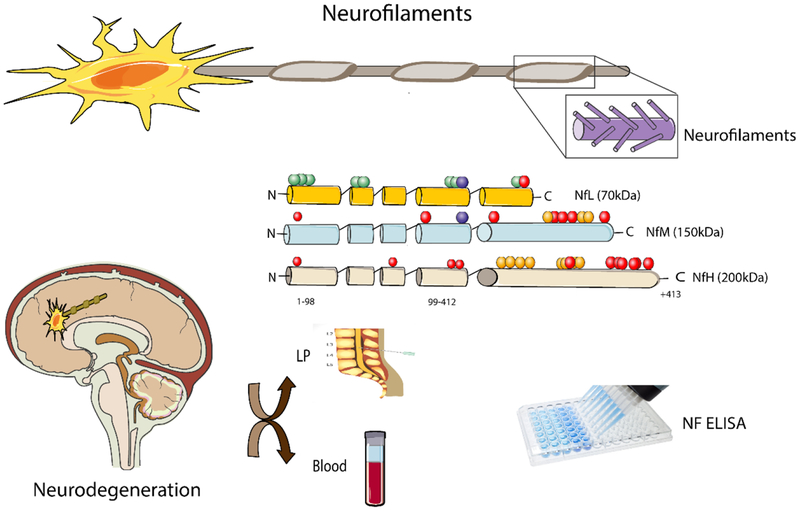
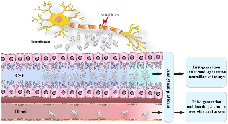
The search for diagnostic and prognostic biomarkers for neurodegenerative conditions is of high importance, since these disorders may present difficulties in differential diagnosis. Biomarkers with high sensitivity and specificity are required. Neurofilament light chain (NfL) is a unique biomarker related to axonal damage and neural cell death, which is elevated in a number of neurological disorders, and can be detected in cerebrospinal fluid (CSF), as well as blood, serum, or plasma samples. Although the NfL concentration in CSF is higher than that in blood, blood measurement may be easier in practice due to its lesser invasiveness, reproducibility, and convenience. Many studies have investigated NfL in both CSF and serum/plasma as a potential biomarker of neurodegenerative disorders. Neuroimaging biomarkers can also potentially improve detection of CNS-related disorders at an early stage. Magnetic resonance imaging (MRI) and diffusion tensor imaging (DTI) are sensitive techniques to visualize neuroaxonal loss. Therefore, investigating the combination of NfL levels with indices extracted from MRI and DTI scans could potentially improve diagnosis of CNS-related disorders. This review summarizes the evidence for NfL being a reliable biomarker in the early detection and disease management in several CNS-related disorders. Moreover, we highlight the correlation between MRI and NfL and ask whether they can be combined.

Keywords: Biomarker; Diffusion tensor imaging; Magnetic resonance imaging; Neurodegenerative disorders; Neurofilament light chain.


Figures

Figure 1.
Figure 1.. A schematic of different types of Nfs and their detection in CNS related disorders.
Nfs act as neurotransmitter. These neurotransmitters are categorized based on their molecular weight. Different types of Nfs could be released in the CSF, serum and plasma. When occurred an injury an increased levels of Nfs will release in various body fluids including CSF, serum and plasma which could be detected by various techniques such as ELISA. NfL: NF light; NfM: NF medium; NfH: NF heavy; ELISA: Enzyme-linked immunosorbent assay
Figure 2.
Figure 2.
A schematic representation of normal and ALS nerve cell. Injured motor neurons in the spinal cord and brain are ALS pathogenesis characteristic. The degenerated neurons are not capable of sending the impulses crucial for movement to the muscle fibers.
Figure 3.
Figure 3.
A schematic showing different events in the pathogenesis of Alzheimer’s disease. An alteration in tau protein leads to microtubule breakdown in brain cells. A healthy neuron and an affected neuron are shown (A, B). Tau phosphorylation contributes to the formation of neurofibrillary tangles in Alzheimer’s disease. In patients with Alzheimer’s disease, hyperphosphorylation of specific amino acids in the tau protein leads to the proteins dissociating from the microtubules, and forming tau tangles. At the same time extracellular amyloid plaque disturbs the transport structure and leads to starvation of neurons, and ultimately induces cell death.
Figure 4.
Figure 4.. The release of Nf after axonal injury.
When an axon is injured, cytoskeletal proteins, such as neurofilaments, are released into the cerebrospinal fluid and, at lower levels, into the blood. First-generation (immunoblots) and second-generation (ELISA) immunoassays can measure neurofilaments in the CSF but have low sensitivity for detecting in the blood. Third-generation (electrochemiluminescence) and fourth-generation (single-molecule array) methods can detect blood concentrations of neurofilament light and measure subtle longitudinal alterations in healthy controls and in pathological cases.

Similar articles

Cited by

References

    1. Strimbu K, Tavel JA (2010) What are biomarkers? Current opinion in HIV and AIDS 5 (6):463–466. doi:10.1097/COH.0b013e32833ed177 - DOI - PMC - PubMed
    1. Califf RM (2018) Biomarker definitions and their applications. Experimental biology and medicine (Maywood, NJ) 243 (3):213–221. doi:10.1177/1535370217750088 - DOI - PMC - PubMed
    1. Lim J, Yue Z (2015) Neuronal aggregates: formation, clearance, and spreading. Developmental cell 32 (4):491–501. doi:10.1016/j.devcel.2015.02.002 - DOI - PMC - PubMed
    1. Jeromin A, Bowser R (2017) Biomarkers in Neurodegenerative Diseases. Adv Neurobiol 15:491–528. doi: 10.1007/978-3-319-57193-5_20. - DOI - PubMed
    1. Di Battista AP, Buonora JE, Rhind SG, Hutchison MG, Baker AJ, Rizoli SB, Diaz-Arrastia R, Mueller GP (2015) Blood biomarkers in moderate-to-severe traumatic brain injury: potential utility of a multi-marker approach in characterizing outcome. Frontiers in neurology 6:110. doi: 10.3389/fneur.2015.00110. - DOI - PMC - PubMed

MeSH terms