. 2019 Dec;18(6):e13026.
doi: 10.1111/acel.13026. Epub 2019 Aug 5.

Long-term repopulation of aged bone marrow stem cells using young Sca-1 cells promotes aged heart rejuvenation

Affiliations

Long-term repopulation of aged bone marrow stem cells using young Sca-1 cells promotes aged heart rejuvenation

Jiao Li et al. Aging Cell. 2019 Dec.

Abstract

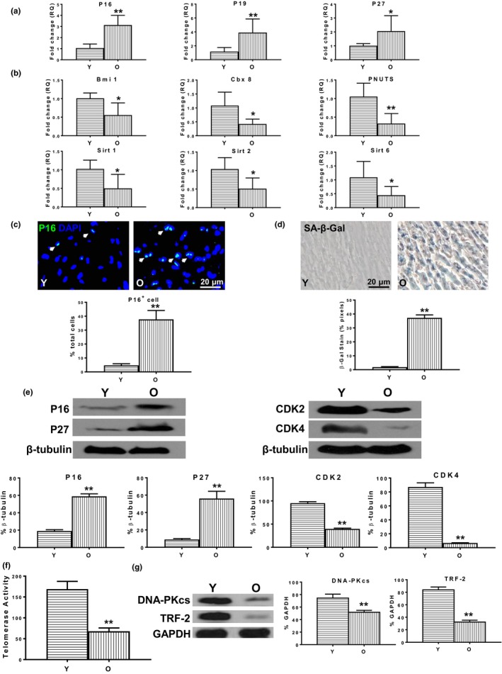
Reduced quantity and quality of stem cells in aged individuals hinders cardiac repair and regeneration after injury. We used young bone marrow (BM) stem cell antigen 1 (Sca-1) cells to reconstitute aged BM and rejuvenate the aged heart, and examined the underlying molecular mechanisms. BM Sca-1+ or Sca-1- cells from young (2-3 months) or aged (18-19 months) GFP transgenic mice were transplanted into lethally irradiated aged mice to generate 4 groups of chimeras: young Sca-1+ , young Sca-1- , old Sca-1+ , and old Sca-1- . Four months later, expression of rejuvenation-related genes (Bmi1, Cbx8, PNUTS, Sirt1, Sirt2, Sirt6) and proteins (CDK2, CDK4) was increased along with telomerase activity and telomerase-related protein (DNA-PKcs, TRF-2) expression, whereas expression of senescence-related genes (p16INK4a , P19ARF , p27Kip1 ) and proteins (p16INK4a , p27Kip1 ) was decreased in Sca-1+ chimeric hearts, especially in the young group. Host cardiac endothelial cells (GFP- CD31+ ) but not cardiomyocytes were the primary cell type rejuvenated by young Sca-1+ cells as shown by improved proliferation, migration, and tubular formation abilities. C-X-C chemokine CXCL12 was the factor most highly expressed in homed donor BM (GFP+ ) cells isolated from young Sca-1+ chimeric hearts. Protein expression of Cxcr4, phospho-Akt, and phospho-FoxO3a in endothelial cells derived from the aged chimeric heart was increased, especially in the young Sca-1+ group. Reconstitution of aged BM with young Sca-1+ cells resulted in effective homing of functional stem cells in the aged heart. These young, regenerative stem cells promoted aged heart rejuvenation through activation of the Cxcl12/Cxcr4 pathway of cardiac endothelial cells.

Keywords: Sca-1; aging; heart; reconstitution; rejuvenation; stem cells.


Conflict of interest statement

The authors have no conflicts of interest to declare.

Figures

Figure 1
Figure 1
Heart senescence increased with aging. The phenotypic changes in cellular senescence were compared in the hearts of young (Y, 2–3 months) and old (O, 22–23 months) wild‐type (C57BL/6) mice. The expression of (a) senescence‐related genes p16INK4a, p19ARF, and p27Kip1 and (b) rejuvenation‐related genes Bmi1, Cbx8, PNUTS, Sirt1, Sirt2, and Sirt6 in O and Y mouse hearts. (c) More p16INK4a+ cells were found in O compared with Y mouse hearts. (d) More senescence‐associated beta galactosidase (SA‐β‐gal)‐positive cells were found in O compared with Y mouse hearts. (e) The protein expression of p16INK4a and p27Kip1 and cyclin‐dependent kinase 2 (CDK2) and CDK4 in O and Y mouse hearts. (f) Telomerase activity and (g) telomerase‐related protein (DNA damage kinases [DNA‐PKcs], telomeric repeat binding factor 2 [TRF‐2]) expression were decreased in O compared with Y mouse hearts. n = 6/group; *p < .05; **p < .01
Figure 2
Figure 2
Cardiac endothelial cells most susceptible to senescence during aging. (a) More p16INK4a and Von Willebrand factor (VWF) double‐positive cells (White arrows) located at the inner lumen side of blood vessels in O mouse hearts. (b) CD31+ endothelial cells were isolated from Y and O wild‐type mouse hearts. The senescence‐associated beta galactosidase activity (SA‐β‐gal, c) was compared in O and Y cardiac endothelial cells. (d) The mRNA expression of p16INK4a, p19ARF, and p27Kip1 and Bmi1, Cbx8, PNUTS, Sirt1, Sirt2, and Sirt6 was compared in O and Y cardiac endothelial cells. (e) The protein expression of p16INK4a, p27Kip1, CDK2, and CDK4 was compared in O and Y cardiac endothelial cells. (f) Telomerase activity and (g) telomerase‐related protein (DNA‐PKcs, TRF‐2) expression were compared in O and Y cardiac endothelial cells. n = 6/group; **p < .01
Figure 3
Figure 3
Young BM Sca‐1 cells decreased recipient cardiac endothelial cell senescence after BM reconstitution. BM Sca‐1+ or Sca‐1 cells (2 × 106) from young (Y, 2–3 months) or old (O, 18–19 months) GFP transgenic mice were transplanted into lethally irradiated (9.5 Gy) O mice to generate 4 groups of chimeras: Y Sca‐1+ (YS+), Y Sca‐1 (YS), O Sca‐1+ (OS+), and O Sca‐1 (OS), respectively. (a) Four months after BM reconstitution, recipient cardiac endothelial cells were isolated from reconstituted mouse hearts using FACS and immunomagnetic bead sorting for GFPCD31+ cells. The senescence‐associated beta galactosidase activity (SA‐β‐gal, b) was compared in recipient cardiac endothelial cells among the 4 chimeric groups. The gene expression of (c) p16INK4a, p19ARF, and p27Kip1 and (d) Bmi1, Cbx8, PNUTS, Sirt1, Sirt2, and Sirt6 was compared among the Y and O and the 4 chimeric groups. The protein expression of (e) p16INK4a and p27Kip1 and (f) CDK2 and CDK4 was compared among the four chimeric groups. (g) Telomerase activity and (h) telomerase‐related protein (DNA‐PKcs, TRF‐2) expression were compared among the 4 chimeric groups. n = 6/group; **p < .01 YS+ vs. YS, OS+ , or OS; *p < .05 YS+ vs. YS, OS+ , or OS; ## p < .01 OS+ vs. OS, #p < .05 OS+ vs. OS
Figure 4
Figure 4
Young BM Sca‐1 cells decreased global senescence of aged recipient hearts. (a) Survival rate of the 4 chimeric groups 4 months after BM reconstitution. (b) p16INK4a+ cells and (c) gene expression of p16INK4a, p19ARF, p27Kip1 Bmi1, Cbx8, PNUTS, Sirt1, Sirt2, and Sirt6 among the Y, O, and the 4 chimeric groups. Protein expression of (d) p16INK4a and p27Kip1 and (e) CDK2 and CDK4. (f) Telomerase activity and (g) telomerase‐related protein (DNA‐PKcs, TRF‐2) in the 4 chimeric hearts. n = 6/group; **p < .01 YS+ vs. other group; ## p < .01 OS+ vs. OS; #p < .05 OS+ vs. OS
Figure 5
Figure 5
Expression profile of growth factors in cardiac homed donor BM cells. Four months after BM reconstitution, myocardial infarction (MI) was induced and donor BM cells (GFP+) were isolated from chimeric hearts at baseline (control without MI) and at 3 days post‐MI. (a) Growth factor qPCR array to profile 84 different growth factors in control groups. Eleven factors were differentially expressed in the homed donor BM cells from the four chimeric groups. The 11 factors were further validated by real‐time qPCR at control as well as at 3 days post‐MI for the4 chimeric groups. These 11 factors were clustered into 5 functional subcategories: (b) cell differentiation regulators (Bmp8a, Bmp5, Lep), (c) apoptosis regulators (Gdf5), (d) angiogenic growth factors (Fgf6, Vegfa), (e) development controllers (Cxcl12, Gdnf, Nodal), and (f) others (IL2, and Tdgf1). n = 3/group for the growth factor qPCR array; n = 6/group for the other experiments; **p < .01 YS+ vs. other group; ## p < .01 OS+ vs. OS; # p < .05 OS+ vs. OS
Figure 6
Figure 6
Young BM Sca‐1 cells decreased senescence of aged recipient cardiac endothelial cells through Cxcl12/Cxcr4 pathway. (a) Gene and (b) protein expression of Cxcr4 was compared in the Y and O wild‐type mouse hearts. Four months after BM reconstitution, recipient cardiac endothelial cells were isolated from reconstituted mouse hearts. (c) Gene and (d) protein expression of Cxcr4 and (e) phosphorylated (P−) and total (T−) AKT, P− and T−, as well as cytoplasmic (C−) and nucleus (N−) forkhead box O3a (FoxO3a). (f) Ratios of P‐AKT/T‐AKT, P‐Fox3a/T‐Fox3a, nuclear (N)‐FoxO3a/N‐PCNA, and cytoplasmic (C)‐FoxO3a/C‐β‐tubulin in recipient cardiac endothelial cells from the four chimeric hearts. n = 6/group; **p < .01 Y vs. O; YS+ vs. other group; ## p < .01 OS+ vs. OS

References

    1. Brunet, A. , Bonni, A. , Zigmond, M. J. , Lin, M. Z. , Juo, P. , Hu, L. S. , … Greenberg, M. E. (1999). Akt promotes cell survival by phosphorylating and inhibiting a Forkhead transcription factor. Cell, 96(6), 857–868. - PubMed
    1. Camici, G. G. , Savarese, G. , Akhmedov, A. , & Luscher, T. F. (2015). Molecular mechanism of endothelial and vascular aging: Implications for cardiovascular disease. European Heart Journal, 36(48), 3392–3403. 10.1093/eurheartj/ehv587 - DOI - PubMed
    1. Capogrossi, M. C. (2004). Cardiac stem cells fail with aging: A new mechanism for the age‐dependent decline in cardiac function. Circulation Research, 94(4), 411–413. 10.1161/01.RES.0000122070.37999.1B - DOI - PubMed
    1. Chen, S. , Lin, J. , Matsuguchi, T. , Blackburn, E. , Yeh, F. , Best, L. G. , … Zhao, J. (2014). Short leukocyte telomere length predicts incidence and progression of carotid atherosclerosis in American Indians: The Strong Heart Family Study. Aging (Albany NY), 6(5), 414–427. 10.18632/aging.100671 - DOI - PMC - PubMed
    1. Cho, R. H. , Sieburg, H. B. , & Muller‐Sieburg, C. E. (2008). A new mechanism for the aging of hematopoietic stem cells: Aging changes the clonal composition of the stem cell compartment but not individual stem cells. Blood, 111(12), 5553–5561. 10.1182/blood-2007-11-123547 - DOI - PMC - PubMed

Publication types