. 2019 Aug 3;2019(1):19-0003.
doi: 10.1530/EDM-19-0003.

A rare ophthalmic condition associated with primary hyperparathyroidism (PHPT): sclerochoroidal calcification (SC)

Affiliations

A rare ophthalmic condition associated with primary hyperparathyroidism (PHPT): sclerochoroidal calcification (SC)

Mona Abouzaid et al. Endocrinol Diabetes Metab Case Rep. .

Abstract

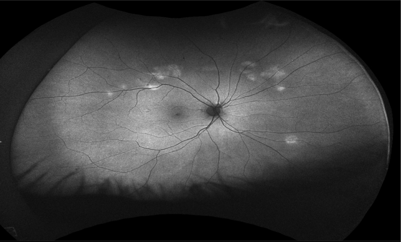
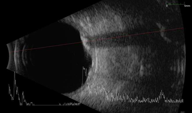
Summary: An 82-year-old male with a proven diagnosis of primary hyperparathyroidism (PHPT) was found to have bilateral changes in the fundi during a routine eye examination which were consistent with SC. In this report, we discuss the link between SC and PHPT and question the need for prospective observational studies to establish the true association between these conditions. Though screening PHPT patients for SC might not be justified/warranted given the benign course of the latter, patients with SC need to be assessed for PHPT, as the former may be the first clue to an underlying treatable systemic disease.

Learning points: Sclerochoroidal calcifications (SCs), though rare and harmless, could be associated with an underlying systemic disease, such as primary hyperparathyroidism (PHPT). Biochemical screening for hypercalcaemia is a simple, cheap and widely available tool that could facilitate an identification of undiagnosed PHPT in patients with SC. A joint care by endocrinologists and ophthalmologists is warranted for those patients, as thorough investigations and long-term follow-up plans are crucial.


Figures

Figure 1
Figure 1
Right eye fundi image.
Figure 2
Figure 2
Left eye fundi image.
Figure 3
Figure 3
Auto fluorescence photographs of the right fundi showed areas of hyperauto fluorescence.
Figure 4
Figure 4
Auto fluorescence photographs of the left fundi showed areas of hyperauto fluorescence.
Figure 5
Figure 5
Ultrasound B-scan of both eyes shows multiple hyper echoic deposits in the posterior ocular coats, persisting at low gain.
Figure 6
Figure 6
CT scan of the orbit shows bilateral sclerochoroidal calcification not involving the extraocular muscles.
Figure 7
Figure 7
Optical coherence tomography (OCT) scanning through an area of calcification at the level of the supratemporal area of the right eye.

References

    1. Lim JI, Goldberg MF. Idiopathic sclerochoroidal calcification: case report. Archives of Ophthalmology 1989. 107 . (10.1001/archopht.1989.01070020188015) - DOI - PubMed
    1. Schachat AP, Robertson DM, Mieler WF, Schwartz D, Augsburger JJ, Schatz H, Gass JD. Sclerochoroidal calcification. Archives of Ophthalmology 1992. 110 . (10.1001/archopht.1992.01080140052025) - DOI - PubMed
    1. Pepe J, Cipriani C, Sonato C, Raimo O, Biamonte F, Minisola S. Cardiovascular manifestations of primary hyperparathyroidism: a narrative review. European Journal of Endocrinology 2017. 177 R297–R308. (10.1530/EJE-17-0485) - DOI - PubMed
    1. Shields CL, Hasanreisoglu M, Saktanasate J, Shields PW, Seibel I, Shields JA. SC: clinical features, outcomes and relationship with hypercalcaemia and parathyroid adenoma in 179 eyes. Retina 2015. 35 . (10.1097/IAE.0000000000000450) - DOI - PubMed
    1. Goldstein BG, Miller J. Metastatic calcification of the choroid in a patient with primary hyperparathyroidism. Retina 1982. 2 . (10.1097/00006982-198200220-00003) - DOI - PubMed