Review
. 2019 Aug 5;50(3):264-282.
doi: 10.1016/j.devcel.2019.06.003.

The Hippo Signaling Pathway in Development and Disease

Affiliations
Review

The Hippo Signaling Pathway in Development and Disease

Yonggang Zheng et al. Dev Cell. .

Abstract

The Hippo signaling pathway regulates diverse physiological processes, and its dysfunction has been implicated in an increasing number of human diseases, including cancer. Here, we provide an updated review of the Hippo pathway; discuss its roles in development, homeostasis, regeneration, and diseases; and highlight outstanding questions for future investigation and opportunities for Hippo-targeted therapies.

Keywords: NF2; TAZ; TEAD; YAP; actomyosin; adherens junction; cancer; cell polarity; cytoskeleton; growth control; hippo signaling; mechanotransduction; merlin; neurofibromatosis; oncogene; regeneration; scalloped; spectrin; tissue homeostasis; tumor suppressor; yorkie.


Figures

Figure 1.
Figure 1.. Kinase Activation Mechanisms in the Hippo Kinase Cascade
Hpo/MST is activated by Tao-1/TAOK-mediated phosphorylation or trans-autophosphorylation of its activation loop site (blue circles). Sav/SAV1 forms a heterotetramer with Hpo/MST to facilitate Hpo/MST activation and localization to the plasma membrane. Activated Hpo/MST then phosphorylates multiple sites (yellow circles) in its linker region. Binding of these phosphorylation sites by Mats/MOB1 helps recruit Wts/LATS to Hpo/MST. Hpo/MST then phosphorylates the HM of Wts/LATS to promote Wts/LATS autophosphorylation and activation. MAP4Ks function redundantly with Hpo/MST to phosphorylate the HM of Wts/LATS leading to its activation. Conversely, the linker phosphorylation sites of Hpo/MST recruit the STRIPAK PP2A phosphatase complex to dephosphorylate and inactivate Hpo/MST, therefore creating a negative feedback to restrict Hpo/MST activity. Upstream regulators such as KIBRA and Mer/NF2 facilitate the kinase cascade by recruiting Wts/LATS to the plasma membrane for its activation by Hpo/MST.
Figure 2.
Figure 2.. The Transcriptional Machinery of the Hippo Signaling Pathway
When Hippo signaling is low, Yki/YAP/TAZ enter the nucleus, where they use their N-terminal homology (NH) domain to interact with the C-terminal domain of the DNA-binding transcriptional factor Sd/TEAD. The DNA-binding TEA domain of Sd/TEAD targets the Yki/YAP/TAZ-Sd/TEAD complex to Hippo Responsive Elements (HREs) of target genes, where Yki/YAP/TAZ activate target gene transcription by recruiting multiple transcriptional complexes, including the Trr (MLL3/4 in mammals) H3K4 methyltranferase complex (through direct binding of its NcoA6 subunit), the ATP-dependent SWI/SNF chromatin remodeling complex, and the Mediator complex. When Hippo signaling is high, Yki/YAP/TAZ are sequestered in the cytoplasm and Sd/TEAD instead interacts with Tgi/VGLL4 and other unknown corepressors to repress target gene transcription.
Figure 3.
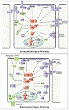
Figure 3.. Upstream Regulators of the Hippo Pathway in Drosophila and Mammals
The signaling diagram illustrates the core kinase cascade (shown within the dashed-line box) and the upstream regulators of the Hippo pathway in both Drosophila and mammals. The upstream regulators link diverse signals such as cell polarity (the Crb/CRB3 complex, the aPKC-PAR complex, and the SCRIB group proteins Scrib, Lgl, and Dlg), cell junctions (adherens junctions in Drosophila and mammals, tight junctions, and focal adhesions in mammals), cytoskeleton (F-actin and spectrin), mechanical forces, GPCR ligands, and some stress signals to the core kinase cascade. The core components of the Hippo kinase cascade are highlighted in red, while the upstream regulators that promote or antagonize Hippo signaling are indicated by purple or green, respectively. Arrowed or blunted ends indicate activation or inhibition, respectively.
Figure 4.
Figure 4.. Hippo Pathway Mutations in Human Diseases
Mutations of Hippo pathway components have been identified in many human diseases, including cancer (highlighted in blue dialogue balloons) and other non-cancer diseases (highlighted in yellow dialogue balloons). The dialogue balloons also point to the specific Hippo pathway components whose mutations are associated with the indicated diseases.

References

    1. Antonescu CR, Le Loarer F, Mosquera JM, Sboner A, Zhang L, Chen CL, Chen HW, Pathan N, Krausz T, Dickson BC, et al. (2013). Novel YAP1-TFE3 fusion defines a distinct subset of epithelioid hemangioendothelioma. Genes Chromosomes Cancer 52, 775–784. - PMC - PubMed
    1. Aragona M, Panciera T, Manfrin A, Giulitti S, Michielin F, Elvassore N, Dupont S, and Piccolo S (2013). A mechanical checkpoint controls multicellular growth through YAP/TAZ regulation by actin-processing factors. Cell 154, 1047–1059. - PubMed
    1. Azzolin L, Panciera T, Soligo S, Enzo E, Bicciato S, Dupont S, Bresolin S, Frasson C, Basso G, Guzzardo V, et al. (2014). YAP/TAZ incorporation in the beta-catenin destruction complex orchestrates the Wnt response. Cell 158, 157–170. - PubMed
    1. Bae SJ, Ni L, Osinski A, Tomchick DR, Brautigam CA, and Luo X (2017). SAV1 promotes Hippo kinase activation through antagonizing the PP2A phosphatase STRIPAK. ELife 6, e30278. - PMC - PubMed
    1. Barolo S, and Posakony JW (2002). Three habits of highly effective signaling pathways: principles of transcriptional control by developmental cell signaling. Genes Dev. 16, 1167–1181. - PubMed

Publication types

MeSH terms