. 2019 Sep:26:101287.
doi: 10.1016/j.redox.2019.101287. Epub 2019 Jul 27.

Therapeutic contribution of melatonin to the treatment of septic cardiomyopathy: A novel mechanism linking Ripk3-modified mitochondrial performance and endoplasmic reticulum function

Affiliations

Therapeutic contribution of melatonin to the treatment of septic cardiomyopathy: A novel mechanism linking Ripk3-modified mitochondrial performance and endoplasmic reticulum function

Jiankai Zhong et al. Redox Biol. 2019 Sep.

Abstract

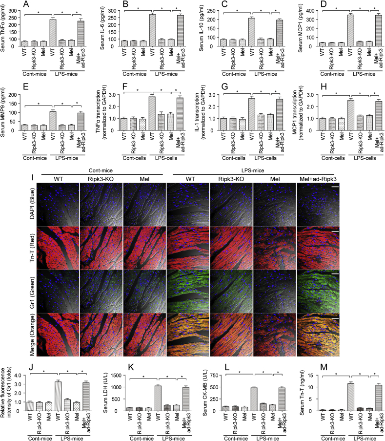
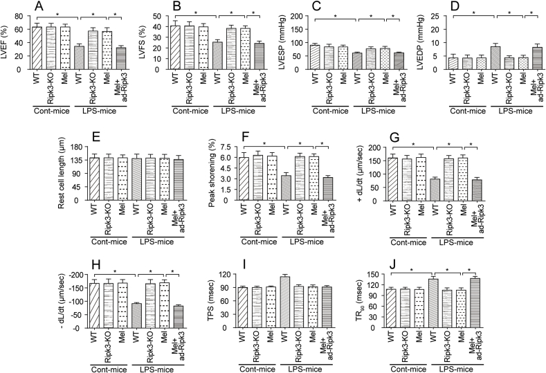
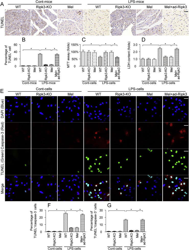
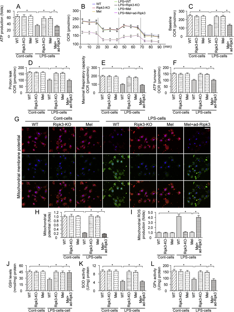
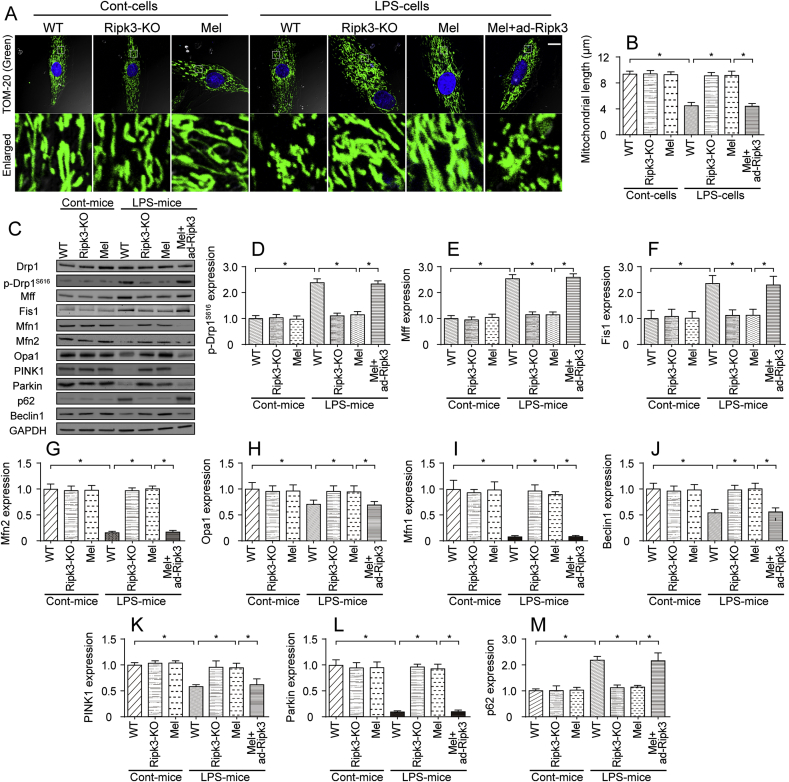
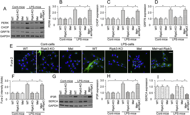
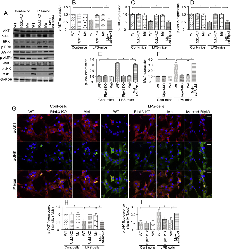
The basic pathophysiological mechanisms underlying septic cardiomyopathy have not yet been completely clarified. Disease-specific treatments are lacking, and care is still based on supportive modalities. The aim of our study was to assess the protective effects of melatonin on septic cardiomyopathy, with a focus on the interactions between receptor-interacting protein kinase 3 (Ripk3), the mitochondria, endoplasmic reticulum (ER) and cytoskeletal degradation in cardiomyocytes. Ripk3 expression was increased in heart samples challenged with LPS, followed by myocardial inflammation, cardiac dysfunction, myocardial breakdown and cardiomyocyte death. The melatonin treatment attenuated septic myocardial injury in a comparable manner to the genetic depletion of Ripk3. Molecular investigations revealed that Ripk3 intimately regulated mitochondrial function, ER stress, cytoskeletal homeostasis and cardioprotective signaling pathways. Melatonin-mediated inhibition of Ripk3 improved mitochondrial bioenergetics, reduced mitochondria-initiated oxidative damage, sustained mitochondrial dynamics, ameliorated ER stress, normalized calcium recycling, and activated cardioprotective signaling pathways (including AKT, ERK and AMPK) in cardiomyocytes. Interestingly, Ripk3 overexpression mediated resistance to melatonin therapy following the infection of LPS-treated hearts with an adenovirus expressing Ripk3. Altogether, our findings identify Ripk3 upregulation as a novel risk factor for the development of sepsis-related myocardial injury, and melatonin restores the physiological functions of the mitochondria, ER, contractile cytoskeleton and cardioprotective signaling pathways. Additionally, our data also reveal a new, potentially therapeutic mechanism by which melatonin protects the heart from sepsis-mediated dysfunction, possibly by targeting Ripk3.

Keywords: Cardioprotective signaling pathways; ER stress; Melatonin; Mitochondrial injury; Ripk3; Septic cardiomyopathy.


Figures

Fig. 1
Fig. 1
Melatonin attenuates sepsis-related myocardial inflammation and injury. A-E. LPS was injected into WT and Ripk3-knockout (Ripk3-KO) mice to establish the septic cardiomyopathy model. Then, melatonin was used to prevent LPS-mediated sepsis. Adenovirus-loaded Ripk3 (ad-Ripk3) was injected into melatonin-treated hearts to reverse the suppression of Ripk3 expression in melatonin-treated mice. Subsequently, blood was isolated and ELISAs were used to detect the alterations in TNFα, IL-6, IL-10, MCP-1 and MMP9 levels. F–H. After LPS-mediated septic cardiomyopathy, qPCR was used to measure the levels of mRNAs encoding inflammatory factors, such as TNFα, IL-1 and MCP1. I-J. Heart tissues were collected and then immunofluorescence staining was performed to analyze the accumulation of Gr-1-positive cells in the myocardium. Tn-T was used to label the myocardial fibers. K-M. After the LPS injection, the concentrations of the cardiac damage markers LDH, CK-MB and Tn-T were determined using ELISAs. *p < 0.05. Bar: 120 μm.
Fig. 2
Fig. 2
LPS-mediated septic cardiomyopathy could be improved by melatonin. A-D. Cardiac function was measured using echocardiography. Then, cardiac systolic and diastolic functions were calculated. Melatonin was injected into the LPS-treated hearts. Adenovirus-loaded Ripk3 (ad-Ripk3) was injected into melatonin-treated hearts to reverse Ripk3. E-J. Cardiomyocyte mechanical parameters were monitored using a VEVO 2100 high-resolution imaging system. Cardiomyocytes were isolated from LPS-treated mice and melatonin-treated mice. Then, the resting cell length, peak shortening, time-to-90% relengthening (TR90), time-to-shortening (TPS), the maximal velocity of shortening (+dL/dt) and the maximal velocity of relengthening (-dL/dt) were determined. *p < 0.05.
Fig. 3
Fig. 3
Melatonin sustained the myocardial contractile cytoskeleton in response to LPS-induced myocardial injury. A-C. Immunofluorescence staining was used to observe the alterations in the cardiac contractile cytoskeleton in vitro. A myosin antibody was used to stain the contractile cytoskeleton. Additionally, tubulin, a component of the noncontractile cytoskeleton in cardiomyocytes, was also stained with a tubulin antibody. Then, the fluorescence intensities of myosin and tubulin were measured. White arrows indicate the myosin degratadion. Bar: 35 μm. D. Electron microscopy was used to observe the alterations in the structure of the cardiac contractile cytoskeleton in vivo. Yellow arrows indicate the Z line in the myocardium. Red arrows indicate the destruction of myocardial fibers. Bar: 2 μm *p < 0.05. (For interpretation of the references to colour in this figure legend, the reader is referred to the Web version of this article.)
Fig. 4
Fig. 4
LPS triggered cardiomyocyte death via necroptosis. A-B. The TUNEL assay was used to evaluate cell death in response to the LPS injection and/or melatonin treatment. The percentage of TUNEL-positive cells was measured. Bar: 180 μm. C. Cardiomyocytes were isolated from mice and then treated with LPS or melatonin. Then, cell viability was evaluated using the MTT assay. D. After treatment with melatonin and/or LPS, the concentrations of LDH were determined using an ELISA, which was used to monitor cardiomyocyte death in response to LPS stress. E-G.In vitro, cardiomyocytes were treated with LPS and/or melatonin. Then, the TUNEL assay and cleaved caspase-3 immunofluorescence staining were conducted. The percentage of TUNEL+/cleaved caspase-3+ cardiomyocytes was counted to determine the number of apoptotic cells. Additionally, the number of TUNEL+/cleaved caspase-3- cardiomyocytes was calculated as the necroptotic cells. Bar: 75 μm *p < 0.05.
Fig. 5
Fig. 5
Mitochondrial bioenergetics were negatively modulated by melatonin through a reduction in Ripk3 expression. A. Cardiomyocytes were isolated from WT and Ripk3-KO mice and then treated with LPS. Subsequently, ATP production was measured to reflect mitochondrial energy metabolism. B–F. Mitochondrial respiratory function was determined by analyzing the mitochondrial oxygen consumption rate (OCR). The baseline OCR, proton leak OCR, maximal respiratory capacity OCR, and ATP turnover OCR were determined. G-H. Mitochondrial membrane potential was measured via JC-1 probe. I. Cardiomyocytes were isolated from WT and Ripk3-KO mice. Then, mitochondrial ROS were stained with MitoSOX red, a mitochondrial superoxide indicator, and mitochondrial ROS production was analyzed using flow cytometry. J-L. Cardiomyocytes were lysed and then ELISAs were used to determine changes in the levels of the antioxidants GSH, GOD and GPx. *p < 0.05. (For interpretation of the references to colour in this figure legend, the reader is referred to the Web version of this article.)
Fig. 6
Fig. 6
Mitochondrial dynamics were modified by melatonin in a manner dependent on Ripk3 repression. A-B. Mitochondrial morphology was determined using immunofluorescence staining with a Tom-20 antibody. In response to the LPS treatment, the mitochondria were divided into several fragments, and these alterations were reversed by melatonin. Then, the mitochondrial length was measured in different groups. Bar: 20 μm. C-M. Mitochondrial fission factors, mitochondrial fusion parameters, and mitophagy markers were determined using western blotting. Drp1 and Mff were the mitochondrial fission factors. Mfn2 and Opa1 were the mitochondrial fusion parameters. Beclin1 and Parkin were the mitophagy markers. *p < 0.05.
Fig. 7
Fig. 7
LPS-induced ER stress and cytoplasmic calcium overloading were inhibited by melatonin. A-D. Cardiomyocytes were isolated from WT and Ripk3-KO mice. Then, the levels of proteins related to ER stress, such as PERK, CHOP, and GRP78, were determined using western blotting. E-F. Cardiomyocyte calcium concentrations was determined using Fura-2AM staining. Subsequently, the fluorescence intensity of calcium was calculated in cells treated with LPS and/or melatonin. Bar: 65 μm. G-I. Proteins were isolated from cardiomyocytes and then western blotting was used to observe the changes in proteins related to ER calcium homeostasis. *p < 0.05.
Fig. 8
Fig. 8
Cardioprotective signaling pathways were modulated by melatonin in the setting of LPS-mediated myocardial injury. A-F. Proteins were isolated from heart tissues and the levels of p-AKT, p-ERK, p-AMPK, p-JNK, and Mst1 were determined. The cardioprotective signaling pathway includes p-AKT, p-ERK, and p-AMPK. However, p-JNK and Mst1 are related to cardiac damage and their levels were also measured using western blotting. G-I. Immunofluorescence staining for p-AKT and p-JNK in cardiomyocytes treated with LPS and melatonin. The immunofluorescence intensity of p-JNK and p-AKT was determined. Bar: 85 μm *p < 0.05.

Similar articles

Cited by

References

    1. Aanhane E., Schulkens I.A., Heusschen R., Castricum K., Leffler H., Griffioen A.W., Thijssen V.L. Different angioregulatory activity of monovalent galectin-9 isoforms. Angiogenesis. 2018;21(3):545–555. - PubMed
    1. Abukar Y., Ramchandra R., Hood S.G., Mckinley M.J., Booth L.C., Yao S.T., May C.N. Increased cardiac sympathetic nerve activity in ovine heart failure is reduced by lesion of the area postrema, but not lamina terminalis. Basic Res. Cardiol. 2018;113(5):35. - PubMed
    1. Abeysuriya R.G., Lockley S.W., Robinson P.A., Postnova S. A unified model of melatonin, 6-sulfatoxymelatonin, and sleep dynamics. J. Pineal Res. 2018;64(4) - PubMed
    1. Angelova P.R., Barilani M., Lovejoy C., Dossena M., Vigano M., Seresini A., Piga D., Gandhi S., Pezzoli G., Abramov A.Y., Lazzari L. Mitochondrial dysfunction in Parkinsonian mesenchymal stem cells impairs differentiation. Redox Biol. 2018;14:474–484. - PMC - PubMed
    1. Frencken J.F., Donker D.W., Spitoni C., Koster-Brouwer M.E., Soliman I.W., Ong D.S.Y., Horn J., Van Der Poll T., Van Klei W.A., Bonten M.J.M., Cremer O.L. Myocardial injury in patients with sepsis and its association with long-term outcome. Circ Cardiovasc Qual Outcomes. 2018;11(2) - PubMed

Publication types

MeSH terms