. 2019 Aug 5;8(8):829.
doi: 10.3390/cells8080829.

Thymic Stromal Lymphopoietin Interferes with the Apoptosis of Human Skin Mast Cells by a Dual Strategy Involving STAT5/Mcl-1 and JNK/Bcl-xL

Affiliations

Thymic Stromal Lymphopoietin Interferes with the Apoptosis of Human Skin Mast Cells by a Dual Strategy Involving STAT5/Mcl-1 and JNK/Bcl-xL

Tarek Hazzan et al. Cells. .

Erratum in

Abstract

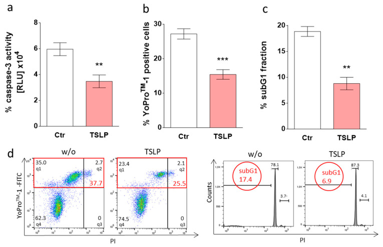
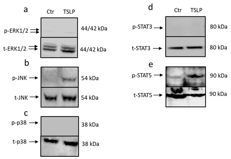
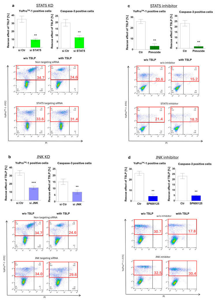
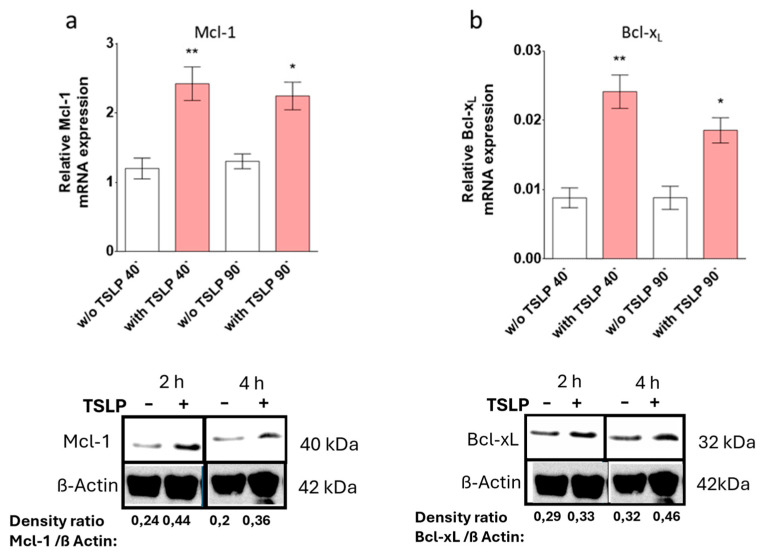
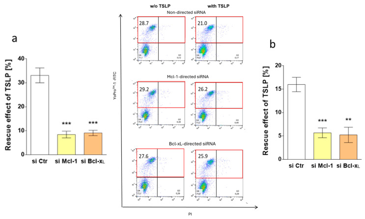
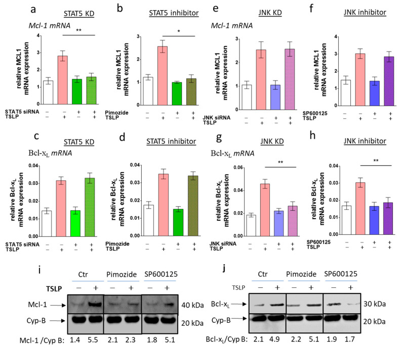
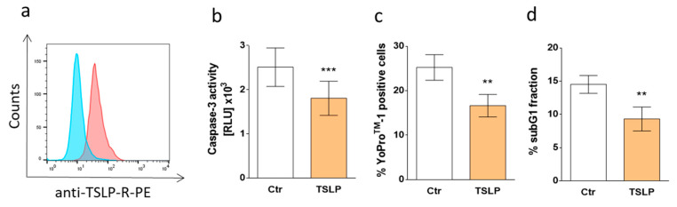
Mast cells (MCs) play critical roles in allergic and inflammatory reactions and contribute to multiple pathologies in the skin, in which they show increased numbers, which frequently correlates with severity. It remains ill-defined how MC accumulation is established by the cutaneous microenvironment, in part because research on human MCs rarely employs MCs matured in the tissue, and extrapolations from other MC subsets have limitations, considering the high level of MC heterogeneity. Thymic stromal lymphopoietin (TSLP)-released by epithelial cells, like keratinocytes, following disturbed homeostasis and inflammation-has attracted much attention, but its impact on skin MCs remains undefined, despite the vast expression of the TSLP receptor by these cells. Using several methods, each detecting a distinct component of the apoptotic process (membrane alterations, DNA degradation, and caspase-3 activity), our study pinpoints TSLP as a novel survival factor of dermal MCs. TSLP confers apoptosis resistance via concomitant activation of the TSLP/ signal transducer and activator of transcription (STAT)-5 / myeloid cell leukemia (Mcl)-1 route and a newly uncovered TSLP/ c-Jun-N-terminal kinase (JNK)/ B-cell lymphoma (Bcl)-xL axis, as evidenced by RNA interference and pharmacological inhibition. Our findings highlight the potential contribution of TSLP to the MC supportive niche of the skin and, vice versa, highlight MCs as crucial responders to TSLP in the context of TSLP-driven disorders.

Keywords: Bcl-xL; JNK; Mcl-1; RNA interference; STAT5; TSLP; apoptosis; mast cells; skin; survival.


Conflict of interest statement

The authors declare no conflict of interest.

Figures

Figure 1
Figure 1
Thymic stromal lymphopoietin (TSLP) counters apoptosis of skin mast cells (MCs) upon grow factor (GF) withdrawal. Skin-derived MCs were kept without or with TSLP (at 7.5 ng/mL) in serum/GF-free medium. (a) Caspase-3 activity as determined after 24 h by Caspase-Glo® 3/7 assay; the results represent the mean ± SEM of six independent experiments. RLU = relative luminescence units (×104). (b,c) Percentage of cells with (b) YoProTM-1 positivity (corresponding to the percentage of early and late apoptotic/necrotic cells combined) after 24 h or (c) fragmented DNA (propidium iodide (PI) staining, corresponding to the percentage of subG1 positive cells) after 48 h. The results represent the mean ± SEM of six (YoProTM-1) or four (PI) independent experiments. (d) Representative flow cytometry dot plots/histograms of (b) (specified in red is the percentage of early and late apoptotic/necrotic cells combined) and (c). The data were analyzed by paired t-test, ** p < 0.01, *** p < 0.001. q—quadrant.
Figure 2
Figure 2
TSLP triggers the activation of signal transducer and activator of transcription (STAT)-5 and c-Jun-N-terminal kinase (JNK). The impact of TSLP treatment (at 7.5 ng/mL) on proximal signaling events after 30 min, as assessed by Western blotting using antibodies for (a) phospho (p) extracellular signal–regulated kinases (ERK)1/2 (n = 3), (b) pJNK, (c) pp38, (d) pSTAT3, and (e) pSTAT5 (shown are representative Western blots out of three independent experiments). The respective total (t) protein antibodies served as loading controls. Undetectable signals were negative also in time-course experiments.
Figure 3
Figure 3
MC maintenance by TSLP critically depends on JNK and STAT5 activation. Impact of (a,c) STAT5 and (b,d) JNK perturbation on TSLP-promoted MC recovery (at 7.5 ng/mL) after 8 h, evaluated by the ratio of YoProTM-1 positivity (corresponding to the percentage of early and late apoptotic/necrotic cells combined) in TSLP-treated versus untreated MCs (described in methods). (a,b) Interference by Accell®-mediated RNAi (48 h prior to TSLP treatment); (c,d) interference by specific inhibitors (STAT5 inhibitor: pimozide, JNK inhibitor: SP600125). Top: the results represent the mean ± SEM of six independent experiments. Bottom: representative flow cytometry dot plots (specified in red is the percentage of early and late apoptotic/necrotic cells combined); w/o—without. The data were analyzed by paired t-test, ** p < 0.01, *** p < 0.001.
Figure 4
Figure 4
TSLP up-regulates Mcl-1 and Bcl-xL. TSLP-induced expression (at 7.5 ng/mL) was studied by (a,b) reverse transcription - quantitative polymerase chain reaction (RT-qPCR) analysis of (a) Mcl-1 and (b) Bcl-xL; normalized to the housekeeping gene Cyclophilin B. The results represent the mean ± SEM of nine independent experiments. The data were analyzed by the one-way Anova test with Tukey’s post-test for multiple comparisons, comparing each treatment (40′ or 90′) with the respective control group; * p < 0.05, ** p < 0.01; and (c,d) Western blot analysis using the indicated antibodies (shown are representative Western blots out of three independent experiments); the anti-β-Actin antibody served as loading control. Densitometry arbitrary units were normalized to the housekeeping protein.
Figure 5
Figure 5
Survival prolongation by TSLP depends on Mcl-1 and Bcl-xL. Impact of Mcl-1 and Bcl-xL knockdown on TSLP-promoted MC recovery (at 7.5 ng/mL), as evaluated by apoptosis reduction in TSLP-treated versus untreated MCs after 8 h. (a) Reduction of YoProTM-1-positivity (corresponding to the percentage of early and late apoptotic/necrotic cells combined) as mean ± SEM of nine independent experiments (left) and representative flow cytometry dot plots (right) (specified in red is the percentage of early and late apoptotic/necrotic cells combined); w/o—without; (b) reduction of caspase-3 activity as mean ± SEM of nine independent experiments. The data were analyzed by paired t-test, ** p < 0.01, *** p < 0.001.
Figure 6
Figure 6
STAT5 perturbation leads to Mcl-1 downregulation, while interference with JNK attenuates Bcl-xL expression. Impact of (ad) STAT5 and (eh) JNK perturbation on TSLP-triggered the expression (at 7.5 ng/mL) of (a,b,e,f) Mcl-1 and (c,d,g,h) Bcl-xL after 40 min, using (a,c,e,g) knockdown by RNAi (48 h prior to the experiment), and (b,d,f,h) pre-incubation with specific inhibitors (STAT5 inhibitor: pimozide, JNK inhibitor: SP600125), evaluated by RT-qPCR analysis (normalized to Cyclophilin B). The results represent the mean ± SEM of five (RNAi) or six (inhibitors) independent experiments. The data were analyzed by the one-way Anova test with Tukey’s post-test for multiple comparisons, * p < 0.05, ** p < 0.01; (i,j) impact of specific inhibitors on the TSLP modulation of (i) Mcl-1 and (j) Bcl-xL by Western blot analysis (shown are representative Western blots out of two independent experiments). Densitometry units were normalized to the housekeeping protein.
Figure 7
Figure 7
(a) Representative flow cytometry histogram of TSLP receptor (TSLPR) surface expression on skin MCs ex vivo (one representative out of four independent stainings is depicted); blue: Isotype control; red: anti-TSLPR. (bd) TSLP counters apoptosis of skin MCs ex vivo. Freshly isolated skin MCs were kept without or with TSLP (at 0.35 ng/mL) in serum/GF-free medium. (b) Caspase-3 activity as determined after 24 h by Caspase-Glo® 3/7 assay; the results represent the mean ± SEM of six independent experiments. RLU = relative luminescence units (×103). (c,d) percentage of cells with (c) YoProTM-1 positivity (corresponding to the percentage of early and late apoptotic/necrotic cells combined) after 48 h or (d) fragmented DNA (PI staining, corresponding to the percentage of subG1 positive cells) after 48 h. The results represent the mean ± SEM of five independent experiments. The data were analyzed by paired t-test, ** p < 0.01, *** p < 0.001.

References

    1. Galli S.J., Tsai M., Piliponsky A.M. The development of allergic inflammation. Nature. 2008;454:445–454. doi: 10.1038/nature07204. - DOI - PMC - PubMed
    1. Kawakami T., Ando T., Kimura M., Wilson B.S., Kawakami Y. Mast cells in atopic dermatitis. Curr. Opin. Immunol. 2009;21:666–678. doi: 10.1016/j.coi.2009.09.006. - DOI - PMC - PubMed
    1. Gilfillan A.M., Austin S.J., Metcalfe D.D. Mast cell biology: Introduction and overview. Adv. Exp. Med. Biol. 2011;716:2–12. doi: 10.1007/978-1-4419-9533-9_1. - DOI - PMC - PubMed
    1. Theoharides T.C., Alysandratos K.D., Angelidou A., Delivanis D.A., Sismanopoulos N., Zhang B., Asadi S., Vasiadi M., Weng Z., Miniati A., et al. Mast cells and inflammation. Biochim. Biophys. Acta Mol. Basis Dis. 2012;1822:21–33. doi: 10.1016/j.bbadis.2010.12.014. - DOI - PMC - PubMed
    1. Galli S.J., Tsai M. IgE and mast cells in allergic disease. Nat. Med. 2012;18:693–704. doi: 10.1038/nm.2755. - DOI - PMC - PubMed

Publication types