Review
. 2019 Aug 5;20(15):3832.
doi: 10.3390/ijms20153832.

A Narrative Role of Vitamin D and Its Receptor: With Current Evidence on the Gastric Tissues

Affiliations
Review

A Narrative Role of Vitamin D and Its Receptor: With Current Evidence on the Gastric Tissues

Shaima Sirajudeen et al. Int J Mol Sci. .

Abstract

Vitamin D is a major steroid hormone that is gaining attention as a therapeutic molecule. Due to the general awareness of its importance for the overall well-being, vitamin D deficiency (VDD) is now recognized as a major health issue. The main reason for VDD is minimal exposure to sunlight. The vitamin D receptor (VDR) is a member of the steroid hormone receptors that induces a cascade of cell signaling to maintain healthy Ca2+ levels that serve to regulate several biological functions. However, the roles of vitamin D and its metabolism in maintaining gastric homeostasis have not yet been completely elucidated. Currently, there is a need to increase the vitamin D status in individuals worldwide as it has been shown to improve musculoskeletal health and reduce the risk of chronic illnesses, including some cancers, autoimmune and infectious diseases, type 2 diabetes mellitus, neurocognitive disorders, and general mortality. The role of vitamin D in gastric homeostasis is crucial and unexplored. This review attempts to elucidate the central role of vitamin D in preserving and maintaining the overall health and homeostasis of the stomach tissue.

Keywords: 1,25-MARRS; 1α,25(OH)2D; cytochrome P450; stomach; vitamin D deficiency; vitamin D epimers.


Conflict of interest statement

The authors declare no conflicts of interest.

Figures

Figure 1
Figure 1
The structure of vitamin D2, vitamin D3, and their precursors. The structural difference between vitamin D2 and D3 is present in their side chains. The side chain of vitamin D3 has a broken ring, while D2 contains a double bond between carbons, 22 and 23, and a methyl group on carbon 24 on the broken ring.
Figure 2
Figure 2
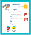
Photobiosynthesis and activation of Vitamin D. 7-dehydrocholesterol in the skin is converted to pre-vitamin D3, upon exposure to sunlight, which contains UVB radiation. Pre-vitamin D3 is converted to vitamin D3 in a heat-dependent process. Vitamin D2 and D3 from the diet are incorporated into chylomicrons and introduced into the circulation. Vitamin D formed in the skin or ingested through diet is stored and released from fat cells. In the serum, vitamin D is circulated while being bound to the vitamin D binding protein, DBP. DBP transports it to the liver where it is converted to 25(OH)D (circulating form) by vitamin D-25-hydroxylase. This form of vitamin D is biologically inactive and is converted in the kidneys to the biologically active form, 1α,25(OH)2D by 25-hydroxyvitamin D-1α- hydroxylase (1α-OHase). The 1α,25(OH)2D binds to the membrane vitamin D receptor (mVDR) or the nuclear vitamin D receptor (nVDR) and elicits specific biological responses.
Figure 3
Figure 3
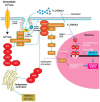
The roles of endocytic proteins in the delivery of 1α,25(OH)2D in the renal cells. The majority of the circulating 25-hydroxyvitamin D is bound to DBP, which is endocytosed via megalin and cubulin-mediated endocytosis. DBP is degraded, and 25(OH)D is either converted to 1α,25(OH)2D in the mitochondria for CYP27B1-mediated bioactivation or is secreted into circulation where it binds to DBP by CYP24A1-mediated inactivation. Cubilin and megalin then return back to the cell surface and the process gets repeated.
Figure 4
Figure 4
A map of the nVDR gene on chromosome 12q12. Blue boxes: Exon 1 (a to f), maroon boxes: Exons 2 to 9, yellow box: 3′ UTR.
Figure 5
Figure 5
The mechanism of action of membrane VDR and nuclear VDR. On binding of the appropriate ligand to mVDR, cellular signal transduction systems that are linked to the membrane receptor get activated, which in turn, trigger the second messengers, resulting in a rapid response. The 1α,25(OH)2D3 binds to the membrane-associated VDR and activates signaling pathways such as PKA and PKC, following which, polyisoprenyl phosphate (PIPP) levels are elevated, thereby triggering the formation of inositol triphosphate (IP3). These signaling pathways help the entry of extracellular calcium into the cells or prompt the release of calcium from intracellular stores in the endoplasmic recticulum (ER). However, binding of 1α,25(OH)2D to the canonical nVDR causes a genomic response by initiating the transcription of targeted genes. Nemere et al. (2004), reported that 1,25-MARRS have a similar affinity for the ligand as that of the nVDR, but, the membrane-associated protein is 6–10 times more abundant in the cells than the nuclear receptor [107]. 1,25-MARRS is usually found associated with caveolin proteins. The intracellular Ca2+ levels are enhanced on binding of 1α,25(OH)2D to 1,25-MARRS. A study in keratinocytes showed that binding of 1α,25(OH)2D3 to the membrane receptor resulted in elevated metabolism of phosphatidylinositol (PI) to phosphatidylinositol triphosphate (PIP3), resulting in increased levels of IP3 in the cells [108,109]. The rise in IP3 were in accordance with a rise in calcium levels, eliciting a rapid response within 2–5 min [50]. Calcium is released from ER storage pools or through the transmembrane trafficking of calcium through the membrane calcium channels [110].
Figure 6
Figure 6
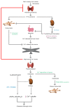
The regulation of mineral homeostasis by parathyroid hormone (PTH) and 1α,25(OH)2D3. The physiological functions of PTH and 1α,25(OH)2D3 are activated when serum calcium levels drop. The hormones act in conjunction with each other and exert coordinated effects on the kidneys, bones, and intestine to increase Ca2+ levels to normal. There is bone resorption, increased calcitriol formation by the kidneys and decreased calcium excretion from urine, and increased Ca2+ absorption by the intestine. Upon achievement of homeostasis, the process is shut down by a negative feedback loop, which is initiated by calcitonin secreted by the thyroid gland. Thus, the combined effect of PTH and 1α,25(OH)2D3 is necessary to maintain mineral homeostasis.

References

    1. Sahota O. Understanding vitamin D deficiency. Age Ageing. 2014;43:589–591. doi: 10.1093/ageing/afu104. - DOI - PMC - PubMed
    1. McKenna M.J., Murray B.F., O’Keane M., Kilbane M.T. Rising trend in vitamin D status from 1993 to 2013: Dual concerns for the future. Endocr. Connect. 2015;4:163–171. doi: 10.1530/EC-15-0037. - DOI - PMC - PubMed
    1. Kuchuk N.O., Pluijm S.M.F., Van Schoor N.M., Looman C.W.N., Smit J.H., Lips P. Relationships of Serum 25-Hydroxyvitamin D to Bone Mineral Density and Serum Parathyroid Hormone and Markers of Bone Turnover in Older Persons. J. Clin. Endocrinol. Metab. 2009;94:1244–1250. doi: 10.1210/jc.2008-1832. - DOI - PubMed
    1. Holick M.F. Vitamin D Deficiency. N. Engl. J. Med. 2007;357:266–281. doi: 10.1056/NEJMra070553. - DOI - PubMed
    1. Chan J., Jaceldo-Siegl K., Fraser G.E. Serum 25-hydroxyvitamin D status of vegetarians, partial vegetarians, and nonvegetarians: The Adventist Health Study-21234. Am. J. Clin. Nutr. 2009;89:1686S–1692S. doi: 10.3945/ajcn.2009.26736X. - DOI - PMC - PubMed