. 2019 Aug 30;9(8):190126.
doi: 10.1098/rsob.190126. Epub 2019 Aug 7.

Mitochondria-hubs for regulating cellular biochemistry: emerging concepts and networks

Affiliations

Mitochondria-hubs for regulating cellular biochemistry: emerging concepts and networks

Alexander J Anderson et al. Open Biol. .

Abstract

Mitochondria are iconic structures in biochemistry and cell biology, traditionally referred to as the powerhouse of the cell due to a central role in energy production. However, modern-day mitochondria are recognized as key players in eukaryotic cell biology and are known to regulate crucial cellular processes, including calcium signalling, cell metabolism and cell death, to name a few. In this review, we will discuss foundational knowledge in mitochondrial biology and provide snapshots of recent advances that showcase how mitochondrial function regulates other cellular responses.

Keywords: metabolism; mitochondria; mitochondrial biogenesis.


Conflict of interest statement

We declare we have no competing interests.

Figures

Figure 1.
Figure 1.
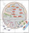
Nuclear-encoded mitochondrial proteins are imported by multi-subunit translocases. Mitochondrial proteins synthesized in the cytosol are imported into mitochondria post-translationally. The TOM complex at the outer membrane serves as a general protein entry gate. hTom40 forms the pore of the translocase, while hTom20, hTom22 and hTom70 function as receptors. hTom22 plays an additional role in the assembly of the complex. hTom5, hTom6 and hTom7, collectively called the small TOMs, regulate the dynamics and assembly of the complex. The TIM22 complex at the inner membrane mediates the import of multi-pass transmembrane proteins into the inner membrane. hTim22 forms the pore through which proteins are inserted, while AGK and hTim29 function as receptors and in complex assembly. The TIM23 complex can translocate precursor proteins into the matrix or the inner membrane. hTim23 and hTim17 form the channel pore, and hTim50 functions as a receptor for precursors. The core complex associates with an import motor that helps to translocate proteins into the matrix in an ATP-dependent manner. The MIA complex mediates the import of soluble intermembrane space proteins by catalysing the formation of disulfide bonds. hMia40 carries out the disulfide bond formation and is anchored to the inner membrane through an interaction with AIF. ALR removes electrons from hMia40 so that it can undergo further rounds of catalysis. The SAM complex of the outer membrane mediates insertion of β-barrel proteins into the outer membrane. hSam50 associates with MTX1 and MTX2. Cristae, the large invaginations of the inner mitochondrial membrane, are stabilized by a multi-subunit complex called MICOS. Mic60 is the core subunit of MICOS, which additionally contains Mic10, Mic13, Mic14, Mic19, Mic25, Mic26 and Mic27. MICOS also associates with the SAM complex at the outer membrane to form a structure known as the mitochondrial intermembrane space bridging complex (MIB).
Figure 2.
Figure 2.
Cellular machineries mediating mitochondrial fission, fusion and formation of contact sites with the endoplasmic reticulum. Mitochondria continuously undergo fission and fusion. Fission is mediated by the GTPase Drp1, which can be recruited to the outer mitochondrial membrane by a variety of receptors, including Mff, Fis1, Mid49 and Mid51. Drp1 at the outer membrane can oligomerize into fibrils that constrict mitochondria to initiate fission. Mitochondrial fusion is initiated by tethering of mitochondria through homotypic interactions between Mfn1 and Mfn2 on opposing mitochondria. Inner membrane fusion is mediated by OPA1, which exists as long and short forms generated through proteolysis. Contact sites between the mitochondria and the endoplasmic reticulum (ER) are established and maintained through protein–protein interactions. Interactions occur between Mfn2 molecules on the ER membrane and the outer mitochondrial membrane, and between VAPB on the ER membrane and RMDN3 on the mitochondrial outer membrane. Interactions also occur between IP3R3, a calcium channel on the ER membrane, and VDAC1 and hTom70 on the mitochondrial outer membrane.
Figure 3.
Figure 3.
Mitochondria coordinate essential metabolic processes. (a) Mitochondria are best known for housing the protein machinery required for generating ATP. When oxygen is available, most cells will generate ATP through oxidative phosphorylation, where electrons harvested through catabolic reactions are used to power ATP synthase. Electrons are obtained through the TCA cycle, which occurs in the matrix and consists of eight enzymatic reactions. Acetyl-CoA is the primary input for the TCA cycle, and can be obtained through metabolism of glucose, fatty acids and amino acids. Electrons extracted during the TCA cycle are loaded onto NAD+ and FAD2+. Electrons are subsequently transferred from NADH and FADH2 onto Complexes I and II of the electron transport chain. Electrons are passed through Complexes III and IV, which transport protons into the intermembrane space. Protons are allowed to flow back into the matrix through ATP synthase (Complex V), which uses the energy of the proton gradient to convert ADP to ATP. (b) Mitochondrial one-carbon (1C) metabolism comprises a series of parallel and reversible reactions which occur in the cytosol and mitochondrial matrix. In proliferating cells, the reaction normally proceeds in a specific direction such that formate produced within mitochondria can be used for biosynthetic processes in the cytosol. Within the mitochondria, THF and serine imported from the cytosol are acted upon sequentially by SHMT2, MTHFD2 and MTHFD1 L to produce formate, which is exported back into the cytosol. Cytosolic MTHFD1 loads formate onto THF to form charged folate intermediates that can be used to synthesize purine and pyrimidine nucleotides. Mitochondrial 1C metabolism is also an important source of glycine. (c) The mitochondrial matrix functions as an important storage site for calcium ions. Mitochondrial calcium uptake often occurs at ER contact sites, where large volumes of Ca2+ can be released through IP3R3. Calcium can pass freely through the outer membrane via VDAC channels and is transported across the intermembrane space and inner membrane through the coordinated function of a MICU1/MICU2 dimer docking to MCU in the inner membrane. Calcium can exit the mitochondrial matrix through LETM1 or SLC8B1 (in exchange for H+ or Na+, respectively) and can cross the outer membrane through VDACs or NCX3.
Figure 4.
Figure 4.
Mitochondria make crucial contributions to diverse cellular processes. (a) The mitochondrial outer membrane is the site of important signalling events during the innate immune response. Detection of viral nucleic acids by Rig-like receptors (RLRs) induces dimerization of MAVS, a protein of the mitochondrial outer membrane. Dimerized MAVS recruits signalling adaptors that initiate downstream activation of IRF3/7 and NF-κB, transcription factors that induce expression of type I interferons and pro-inflammatory cytokines. MAVS is regulated by NLRX1, a protein which downregulates MAVS when localized to the outer membrane, but activates MAVS when at the inner membrane by interacting with Complex III to induce ROS production. Release of mtDNA during infection can also activate the NLRP3 inflammasome. (b) Mitophagy is a process that allows damaged mitochondria to be identified and destroyed. Under normal conditions, PINK1 is imported into mitochondria and degraded by PARL. When mitochondria are damaged, import is impaired and PINK1 accumulates in the TOM complex at the outer membrane. Autophosphorylated and active PINK1 at the outer membrane phosphorylates monoubiquitin molecules on outer membrane proteins, recruiting and activating the E3 ubiquitin ligase Parkin. Activated Parkin synthesizes polyubiquitin chains that recruit autophagy receptors to initiate mitophagy. (c) Mitochondrial proteostatic stress is sensed through the partitioning of the transcription factor ATF5 between the mitochondria and the nucleus. Under normal conditions, ATF5 is imported into and sequestered within mitochondria. If mitochondrial protein import becomes compromised, ATF5 is trafficked into the nucleus, where it upregulates expression of genes that enhance proteostasis. (d) Mitochondria play crucial roles in the initiation of apoptosis. In response to pro-apoptotic stimuli, Bax and Bak oligomerize in the outer membrane to form pores that allow for efflux of apoptogenic proteins (Cytochrome c, Diablo, AIF and Endonuclease G) from the intermembrane space into the cytosol. Cytochrome c binds to Apaf-1 to induce formation of the apoptosome and activation of caspases. Diablo blocks inhibitors of apoptosis (IAPs) which would otherwise mitigate the effect of caspases. AIF and Endonuclease G translocate into the nucleus where they contribute to destruction of the genome.

Similar articles

Cited by

References

    1. Gray MW, Burger G, Lang BF. 1999. Mitochondrial evolution. Science 283, 1476–1481. (10.1126/science.283.5407.1476) - DOI - PubMed
    1. Cogliati S, Enriquez JA, Scorrano L. 2016. Mitochondrial cristae: where beauty meets functionality. Trends Biochem. Sci. 41, 261–273. (10.1016/j.tibs.2016.01.001) - DOI - PubMed
    1. Hoppins S, et al. 2011. A mitochondrial-focused genetic interaction map reveals a scaffold-like complex required for inner membrane organization in mitochondria. J. Cell Biol. 195, 323 (10.1083/jcb.201107053) - DOI - PMC - PubMed
    1. Gödiker J, et al. 2018. QIL1-dependent assembly of MICOS complex–lethal mutation in C19ORF70 resulting in liver disease and severe neurological retardation. J. Hum. Genet. 63, 707–716. (10.1038/s10038-018-0442-y) - DOI - PubMed
    1. Ding C, et al. 2015. Mitofilin and CHCHD6 physically interact with Sam50 to sustain cristae structure. Sci. Rep. 5, 16064 (10.1038/srep16064) - DOI - PMC - PubMed

LinkOut - more resources