

Atherosclerosis and flow: roles of epigenetic modulation in vascular endothelium
- PMID: 31387590
- PMCID: PMC6685237
- DOI: 10.1186/s12929-019-0551-8
Atherosclerosis and flow: roles of epigenetic modulation in vascular endothelium
Abstract
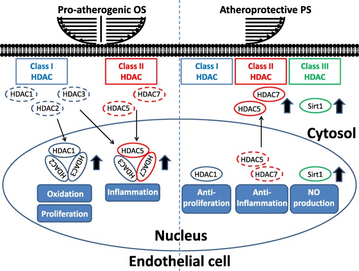
Background: Endothelial cell (EC) dysfunctions, including turnover enrichment, gap junction disruption, inflammation, and oxidation, play vital roles in the initiation of vascular disorders and atherosclerosis. Hemodynamic forces, i.e., atherprotective pulsatile (PS) and pro-atherogenic oscillatory shear stress (OS), can activate mechanotransduction to modulate EC function and dysfunction. This review summarizes current studies aiming to elucidate the roles of epigenetic factors, i.e., histone deacetylases (HDACs), non-coding RNAs, and DNA methyltransferases (DNMTs), in mechanotransduction to modulate hemodynamics-regulated EC function and dysfunction. OS enhances the expression and nuclear accumulation of class I and class II HDACs to induce EC dysfunction, i.e., proliferation, oxidation, and inflammation, whereas PS induces phosphorylation-dependent nuclear export of class II HDACs to inhibit EC dysfunction. PS induces overexpression of the class III HDAC Sirt1 to enhance nitric oxide (NO) production and prevent EC dysfunction. In addition, hemodynamic forces modulate the expression and acetylation of transcription factors, i.e., retinoic acid receptor α and krüppel-like factor-2, to transcriptionally regulate the expression of microRNAs (miRs). OS-modulated miRs, which stimulate proliferative, pro-inflammatory, and oxidative signaling, promote EC dysfunction, whereas PS-regulated miRs, which induce anti-proliferative, anti-inflammatory, and anti-oxidative signaling, inhibit EC dysfunction. PS also modulates the expression of long non-coding RNAs to influence EC function. i.e., turnover, aligmant, and migration. On the other hand, OS enhances the expression of DNMT-1 and -3a to induce EC dysfunction, i.e., proliferation, inflammation, and NO repression.
Conclusion: Overall, epigenetic factors play vital roles in modulating hemodynamic-directed EC dysfunction and vascular disorders, i.e., atherosclerosis. Understanding the detailed mechanisms through which epigenetic factors regulate hemodynamics-directed EC dysfunction and vascular disorders can help us to elucidate the pathogenic mechanisms of atherosclerosis and develop potential therapeutic strategies for atherosclerosis treatment.
Keywords: DNA methyltransferase; Endothelial cell; Epigenetic factor; Hemodynamic force; Histone deacetylase; Non-coding RNA.
Conflict of interest statement
The authors declare that they have no competing interests.
Figures

 : pro-atherogenic OS;
 : atheroprotective PS

References
Publication types
MeSH terms
Substances
Grants and funding
- MOST-108-2320-B-157-001/Ministry of Science and Technology, Taiwan
- MOST-107-2321-B-400-001/Ministry of Science and Technology, Taiwan
- MOST-107-2320-B-157-001/Ministry of Science and Technology, Taiwan
- MOST-108-2633-B-009-001/Ministry of Science and Technology, Taiwan
- 108-1901-01-19-08/108-1901-01-19-07/107-1901-01-19-03/ MOST106-3114-Y-043-021/108-0324-01-19-07/107-0324-01-19-03/106-0324-01-10-07/105-0324-01-10-03/National Health Research Institutes, and Central Government S & T grants, Taiwan
LinkOut - more resources
Full Text Sources
Medical
Miscellaneous
