. 2019 Aug 6;38(1):341.
doi: 10.1186/s13046-019-1349-y.

Depleting PTOV1 sensitizes non-small cell lung cancer cells to chemotherapy through attenuating cancer stem cell traits

Affiliations

Depleting PTOV1 sensitizes non-small cell lung cancer cells to chemotherapy through attenuating cancer stem cell traits

Zhiqiang Wu et al. J Exp Clin Cancer Res. .

Abstract

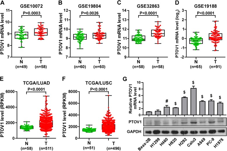
Background: Prostate tumor over expressed gene 1 (PTOV1) has been reported as an oncogene in several human cancers. However, the clinical significance and biological role of PTOV1 remain elusive in non-small cell lung cancer (NSCLC).

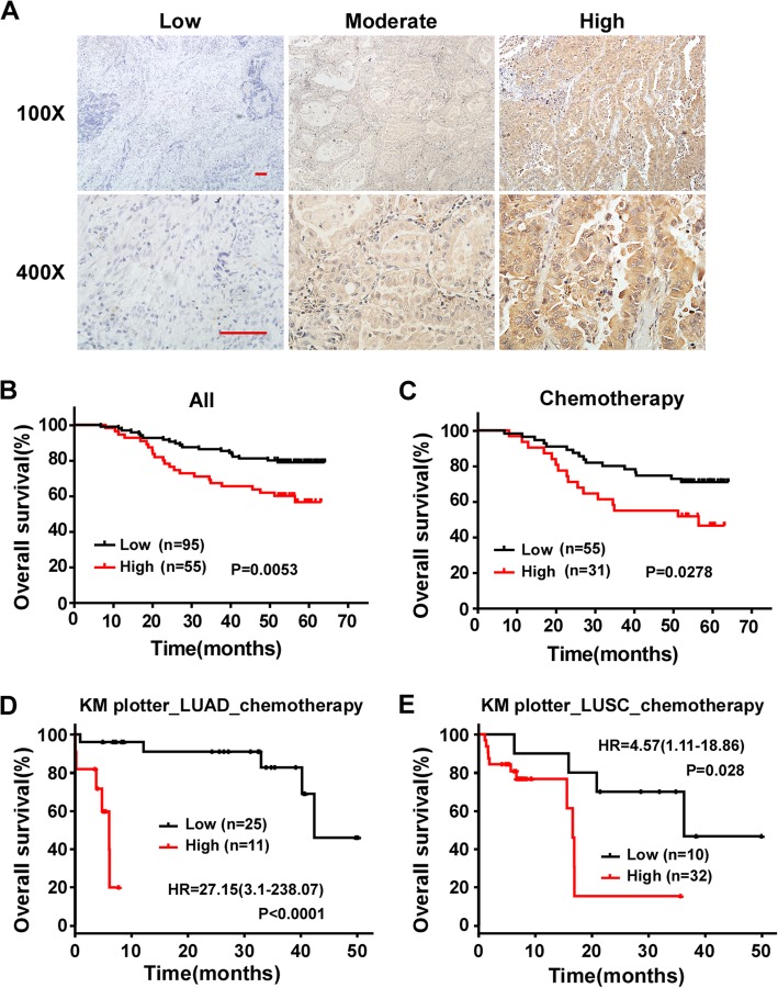
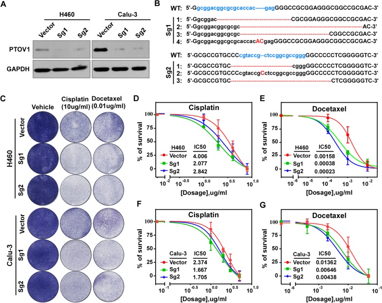
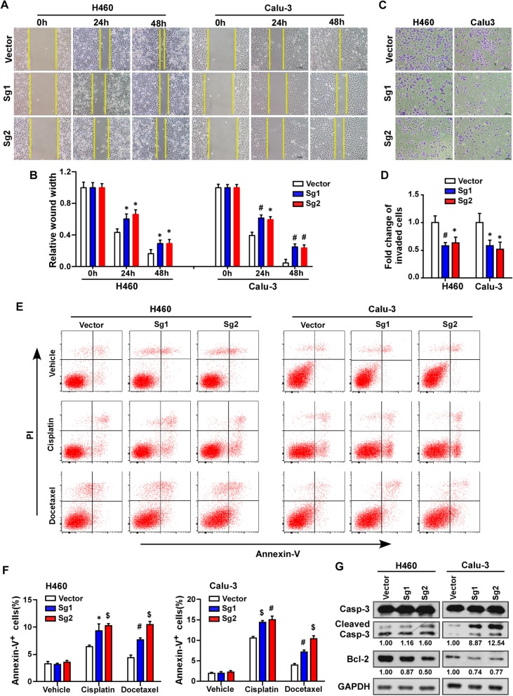
Methods: The Cancer Genome Atlas (TCGA) data and NCBI/GEO data mining, western blotting analysis and immunohistochemistry were employed to characterize the expression of PTOV1 in NSCLC cell lines and tissues. The clinical significance of PTOV1 in NSCLC was studied by immunohistochemistry statistical analysis and Kaplan-Meier Plotter database mining. A series of in-vivo and in-vitro assays, including colony formation, CCK-8 assays, flow cytometry, wound healing, trans-well assay, tumor sphere formation, quantitative PCR, gene set enrichment analysis (GSEA), immunostaining and xenografts tumor model, were performed to demonstrate the effects of PTOV1 on chemosensitivity of NSCLC cells and the underlying mechanisms.

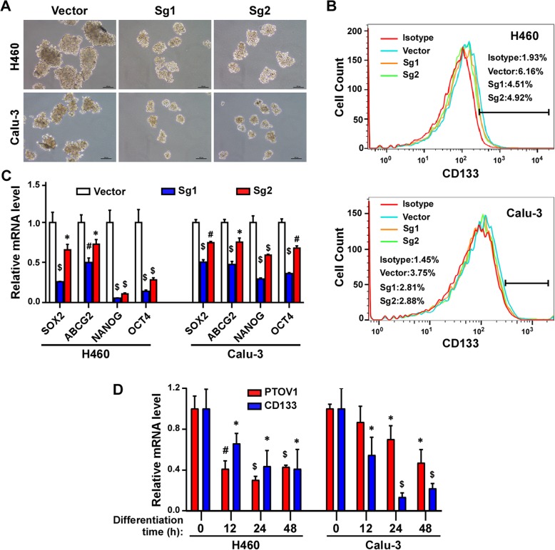
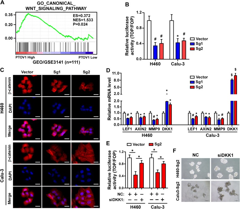
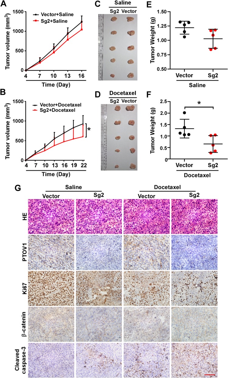
Results: PTOV1 is overexpressed in NSCLC cell lines and tissues. High PTOV1 level indicates a short survival time and poor response to chemotherapy of NSCLC patients. Depleting PTOV1 increased sensitivity to chemotherapy drugs cisplatin and docetaxel by increasing cell apoptosis, inhibiting cell migration and invasion. Our study verified that depleting PTOV1 attenuated cancer stem cell traits through impairing DKK1/β-catenin signaling to enhance chemosensitivity of NSCLC cells.

Conclusion: These results suggest that PTOV1 plays an important role in the development and progression of human NSCLC and PTOV1 may serve as a therapeutic target for NSCLC patients.

Keywords: Cancer stem cell; Chemotherapy; Non-small cell lung cancer; PTOV1; β-Catenin.


Conflict of interest statement

The authors declare that they have no competing interests.

Figures

Fig. 1
Fig. 1
PTOV1 is upregulated in NSCLC. a-d Analysis of the expression of PTOV1 mRNA in NSCLC datasets from the GEO database. e and f Analysis of the expression of PTOV1 mRNA in NSCLC datasets from the TCGA database. g Real-time PCR (upper panel) and western blotting (lower panel) analyses of PTOV1 expression in lung epithelial cell line, Beas-2B, and 8 NSCLC cell lines. N, normal tissue. T, tumor tissue. Error bars represent the mean ± SD form 3 independent experiments. P values are calculated by two-tailed, unpaired t-test. *, P < 0.05; #, P < 0.01; $, P < 0.001
Fig. 2
Fig. 2
High PTOV1 expression correlates with poor prognosis and chemo resistance in NSCLC patients. a Representative pictures of NSCLC tissues with low, modulate and high PTOV1 expression levels analyzed by IHC staining. Scale bars, 50 μm. b and c Kaplan-Meier analysis of overall survival in all NSCLC patients (b) and the subgroup patients received chemotherapy (c). d and e Kaplan-Meier analysis of overall survival in the LUAD (d) and LUSC (e) subgroup patients received chemotherapy by the Kaplan–Meier Plotter online database. P values are calculated by log-rank test
Fig. 3
Fig. 3
Depleting PTOV1 increases sensitivities to cisplatin or docetaxel of NSCLC cells. a Western blotting analyzing the expression of PTOV1 in the indicated cells. b Representative sequencing results of different indels introduced by SgRNAs in pooled PTOV1-Sg1 and –Sg2 cells comparing to the wide type (WT) PTOV1 locus. Blue lowercase letters indicate the target sequences of SgRNA 1 and 2. Red dash lines and capital letters are the indels. c Colony formation of the indicated cells after treatment with cisplatin or docetaxel. d-g IC50s the indicated cells responding to cisplatin or docetaxel determined by CCK-8 assay
Fig. 4
Fig. 4
Depleting PTOV1 inhibits migration and invasion and promotes cell apoptosis. a and b Representative images (a) and quantification (b) of cell migration the indicated cells analyzed by wound healing assay. c and d Representative images (c) and quantification (d) of invade cells analyzed by chamber invasion assay. e and f Representative images (e) and quantification (f) of cell apoptosis of the indicated cells treated with or without drugs analyzed by Annexin V/PI staining. g Western blotting analysis of Caspase-3 (Casp-3), Cleaved Casp-3 and Bcl-2. GAPDH served as a loading control. Error bars represent mean ± SD from least 3 independent experiments. *, P < 0.05; #, P < 0.01; $, P < 0.001, two-tailed, unpaired t-test
Fig. 5
Fig. 5
Depleting PTOV1 impaired stem cell-like properties in NSCLC cells. a Representative images of tumor sphere formation of the indicated cells. b FACS analysis of the portion of CD133+ cells. c Real-time quantitative PCR detecting the expression of the pluripotency factors in the indicated cells. d Real-time quantitative PCR detecting the expression of PTOV1 and CD133 in undifferentiated and serum-induced differentiated cells. Error bars represent the mean ± SD from 3 independent experiments. *, P < 0.05; #, P < 0.01; $, P < 0.001, two-tailed, unpaired t-test
Fig. 6
Fig. 6
Depleting PTOV1 impaired stem cell-like properties via DKK1/β-catenin signaling. a GSEA analysis of the correlation between PTOV1 expression and the GO_ CANONICAL_WNT_SIGNALING_PATHWAY gene signature. b TOP/FOP Flash activity of the indicated cells. c Representative images of immunofluorescence staining of β-catenin in the indicated cells. d Real-time quantitative PCR detecting the expression of LEF1, AXIN2, MMP9and DKK1 in the indicated cells. e TOP/FOP Flash activity of the indicated cells transfected with NC or DKK siRNA. f Representative images of tumor sphere formation of the indicated cells transfected with NC or DKK siRNA. Error bars represent the mean ± SD from 3 independent experiments. *, P < 0.05; #, P < 0.01; $, P < 0.001, two-tailed, unpaired t-test
Fig. 7
Fig. 7
Depleting PTOV1 chemosensitizes NSCLC cells in vivo. a and d Growth curve of the tumor volumes measured on the indicated days. Error bars represent the mean ± SD. *, P < 0.05, one-way ANOVA test. b and e Representative pictures of tumor growth. c and f Quantification of tumor weights. Error bars represent the mean ± SD. *, P < 0.05, two-tailed, unpaired t-test. g Representative pictures of H&E staining and IHC staining of PTOV1, Ki-67, β-catenin and cleaved Caspase-3 in the indicated xenografted tumors. Scale bar, 50 μm

Similar articles

Cited by

References

    1. Torre LA, Bray F, Siegel RL, Ferlay J, Lortet-Tieulent J, Jemal A. Global cancer statistics, 2012. CA Cancer J Clin. 2015;65(2):87–108. doi: 10.3322/caac.21262. - DOI - PubMed
    1. Siegel RL, Miller KD, Jemal A. Cancer statistics, 2016. CA Cancer J Clin. 2016;66(1):7–30. doi: 10.3322/caac.21332. - DOI - PubMed
    1. Arriagada R, Bergman B, Dunant A, Le Chevalier T, Pignon JP, Vansteenkiste J, et al. Cisplatin-based adjuvant chemotherapy in patients with completely resected non-small-cell lung cancer. N Engl J Med. 2004;350(4):351–360. doi: 10.1056/NEJMoa031644. - DOI - PubMed
    1. Winton T, Livingston R, Johnson D, Rigas J, Johnston M, Butts C, et al. Vinorelbine plus cisplatin vs. observation in resected non-small-cell lung cancer. N Engl J Med. 2005;352(25):2589–2597. doi: 10.1056/NEJMoa043623. - DOI - PubMed
    1. Chang A. Chemotherapy, chemoresistance and the changing treatment landscape for NSCLC. Lung Cancer. 2011;71(1):3–10. doi: 10.1016/j.lungcan.2010.08.022. - DOI - PubMed

MeSH terms