. 2019 Sep 2;151(9):1081-1093.
doi: 10.1085/jgp.201912431. Epub 2019 Aug 6.

The central role of the tail in switching off 10S myosin II activity

Affiliations

The central role of the tail in switching off 10S myosin II activity

Shixin Yang et al. J Gen Physiol. .

Abstract

Myosin II is a motor protein with two heads and an extended tail that plays an essential role in cell motility. Its active form is a polymer (myosin filament) that pulls on actin to generate motion. Its inactive form is a monomer with a compact structure (10S sedimentation coefficient), in which the tail is folded and the two heads interact with each other, inhibiting activity. This conformation is thought to function in cells as an energy-conserving form of the molecule suitable for storage as well as transport to sites of filament assembly. The mechanism of inhibition of the compact molecule is not fully understood. We have performed a 3-D reconstruction of negatively stained 10S myosin from smooth muscle in the inhibited state using single-particle analysis. The reconstruction reveals multiple interactions between the tail and the two heads that appear to trap ATP hydrolysis products, block actin binding, hinder head phosphorylation, and prevent filament formation. Blocking these essential features of myosin function could explain the high degree of inhibition of the folded form of myosin thought to underlie its energy-conserving function in cells. The reconstruction also suggests a mechanism for unfolding when myosin is activated by phosphorylation.


Figures

Figure 1.
Figure 1.
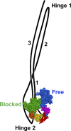
Model of 10S myosin. Blocked head consists of heavy chain (green), ELC (orange), and RLC (yellow). Free head shows heavy chain (blue), ELC (magenta), and RLC (red). The tail is folded into three segments: segment 1 (subfragment 2), originating from the heads and ending at hinge 1; segment 2, starting at hinge 1 and ending at hinge 2; and segment 3, starting at hinge 2 and ending at the tip of the tail. Based on Burgess et al. (2007) and Lee et al. (2018).
Figure 2.
Figure 2.
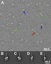
Negatively stained 10S smooth muscle myosin molecules. (A) Two types of structure are visible: monomers, in front and side views (green and blue arrows, respectively), and occasional dimers (red arrow). (B–D) 2-D class averages of right and left views (B and C, respectively; Jung et al. [2008b]); partially rotated view (D); and side view (E; see also Fig. S1).
Figure 3.
Figure 3.
3-D reconstruction of 10S smooth muscle myosin. Electron Microscopy Databank accession code EMD-20084. (A–C) Front, back, and side views, respectively. (D–F) D–F correspond to A–C, with colors indicating different components of the structure. Blocked head is red; free head, green; tail segments 1 and 3, cyan; segment 2, magenta. In E, yellow circle indicates putative location of missing segment 1 density (see Fig. S3); red arrow, start of segment 3. Yellow star in D indicates the second hinge point.
Figure 4.
Figure 4.
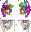
Docking of atomic model of the IHM into the reconstruction. The 2-D crystal model of smooth muscle myosin (PDB ID 1I84; Wendt et al., 2001) was used in the fitting. Molecular dynamics flexible fitting was not used, due to the low resolution. (A–C) Front, back, and side views, respectively, docked with ribbon representation of the atomic model. (D–F) D–F correspond to A–C, but with space-filling model. The atomic model fits well into the reconstruction in all views, except for the narrow part of the lever arm in the blocked head (single α-helix, between the two light chains; red arrow) and part of the ELC in the free head (yellow arrow). The low density in the reconstruction at these points may be due to extension of the stain pool that accumulates in the hole at the center of the IHM (cf. Figs. 2 B and S1) into these low-density regions of protein, obscuring them. Unfilled densities mostly represent portions of the three segments of the tail, for which there is no atomic model. There is also some unfilled density in the blocked head (green arrow) that would be filled by repositioning the flexibly connected SH3 domain (not done with the rigid-body docking procedure; see Materials and methods). In a previous study, molecular dynamics flexible fitting of cryo-imaged tarantula thick filaments demonstrated this point (Yang et al., 2016). Note: C and F rotated in opposite direction from C and F in Fig. 3.
Figure 5.
Figure 5.
Interactions between the tail and heads in 10S myosin. (A) Front view of reconstruction fitted with PDB ID 1I84 shows the interactions between tail segments 2 (magenta) and 3 (cyan) and the blocked head (TB2, TB3, TB4, TB5), between the two heads (BF), and between segment 2 and the other tail segments (the interaction extends upwards from TT1). TB2 is between the SH3 domain (blue) in the blocked head and segment 2, lying underneath. TB3 is between segment 2 and the converter domain (cyan) of the blocked head. TB4 is between segment 2 and the ELC (orange) of the blocked head. TB5 is between segment 3 and the blocked-head RLC. (B) Back view shows the interaction between the free head and the tail (TF1, TF2, TF3), between segments 1 and 3 (unresolved from each other) and the blocked head (TB1), and between tail segments 1 and 3 (starting at TT2). Interaction TF1 is between the actin-binding loop in the free head and the tail, while TF2 is between the free head upper 50K domain and the tail. TF3 may represent merging of tail and blocked head densities and not be a real interaction (see Results). TB1 is an extended interaction between the blocked head and segment 1 and/or segment 3. (C) Magnified view of TB5. Cys108 is represented by a cyan sphere. (D) The view in C is rotated 135° to visualize the geometric relationship between Cys108 and segment 3.
Figure 6.
Figure 6.
Heterogeneity in the reconstruction. 3-D classification using RELION produced six classes of conformation (A–F, classes 1–6, respectively), showing that the 10S myosin molecules are quite heterogeneous, due either to variable staining and/or other factors during specimen preparation or to natural variation in the molecules themselves. (A) The tail density partially disappeared in some regions (red dotted circles; cf. red arrow in C, where it is visible). (B) The tail cannot be explicitly identified in this reconstruction, and the second hinge point (star in C) is missing (blue circle). The reconstructions in C and D showed similar structural features, with both the interacting heads and the tail clearly visible. (E) Segment 2 around the blocked head is not visible, and the second hinge point is absent (blue circle), as in B. (F) The tail wraps around the blocked head, but the density present in C (cyan arrow) and D (cyan circle) is absent.
Figure 7.
Figure 7.
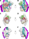
Proposed inhibition mechanism of phosphate release and actin binding in the two heads. (A) Phosphate release in the blocked head is inhibited by interaction of its converter domain (cyan within red ellipse) with segment 2, and in the free head by interaction of its converter domain (cyan within blue ellipse) with the blocked head. (B) Back view, rotated 180° from A. (C and D) Front and back views show actin-binding loop (red ellipse) in the blocked head sterically blocked by the free head, and actin-binding loop (green ellipse) in the free head blocked by interacting with the tail. (E and F) Front and back views showing steric blocking of Ser19 (red sphere) by segment 3 in the blocked-head RLC, but not in the free-head RLC (magenta sphere; see Video 4).

Comment in

References

    1. Alamo L., Wriggers W., Pinto A., Bártoli F., Salazar L., Zhao F.Q., Craig R., and Padrón R.. 2008. Three-dimensional reconstruction of tarantula myosin filaments suggests how phosphorylation may regulate myosin activity. J. Mol. Biol. 384:780–797. 10.1016/j.jmb.2008.10.013 - DOI - PMC - PubMed
    1. Alamo L., Qi D., Wriggers W., Pinto A., Zhu J., Bilbao A., Gillilan R.E., Hu S., and Padrón R.. 2016. Conserved Intramolecular Interactions Maintain Myosin Interacting-Heads Motifs Explaining Tarantula Muscle Super-Relaxed State Structural Basis. J. Mol. Biol. 428:1142–1164. 10.1016/j.jmb.2016.01.027 - DOI - PMC - PubMed
    1. Ankrett R.J., Rowe A.J., Cross R.A., Kendrick-Jones J., and Bagshaw C.R.. 1991. A folded (10 S) conformer of myosin from a striated muscle and its implications for regulation of ATPase activity. J. Mol. Biol. 217:323–335. 10.1016/0022-2836(91)90546-I - DOI - PubMed
    1. Baker T.S., and Johnson J.E.. 1996. Low resolution meets high: towards a resolution continuum from cells to atoms. Curr. Opin. Struct. Biol. 6:585–594. 10.1016/S0959-440X(96)80023-6 - DOI - PubMed
    1. Breckenridge M.T., Dulyaninova N.G., and Egelhoff T.T.. 2009. Multiple regulatory steps control mammalian nonmuscle myosin II assembly in live cells. Mol. Biol. Cell. 20:338–347. 10.1091/mbc.e08-04-0372 - DOI - PMC - PubMed

Publication types

Substances