. 2019 Aug 6;10(4):e01238-19.
doi: 10.1128/mBio.01238-19.

Anopheles Salivary Gland Architecture Shapes Plasmodium Sporozoite Availability for Transmission

Affiliations

Anopheles Salivary Gland Architecture Shapes Plasmodium Sporozoite Availability for Transmission

Michael B Wells et al. mBio. .

Abstract

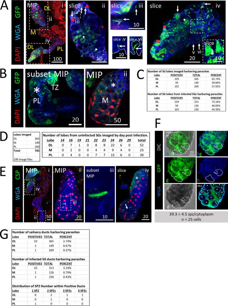
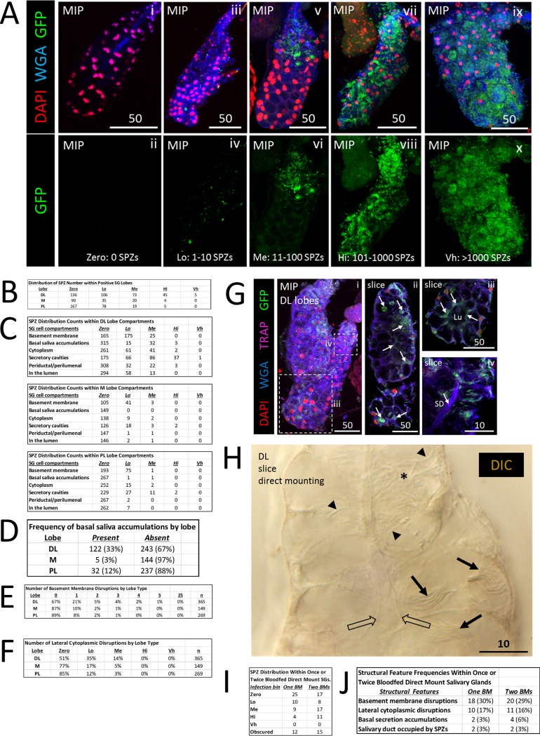
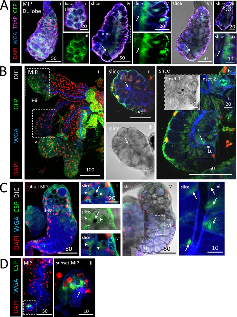
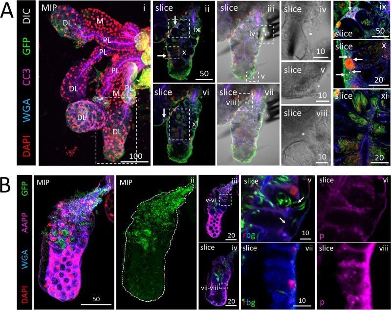
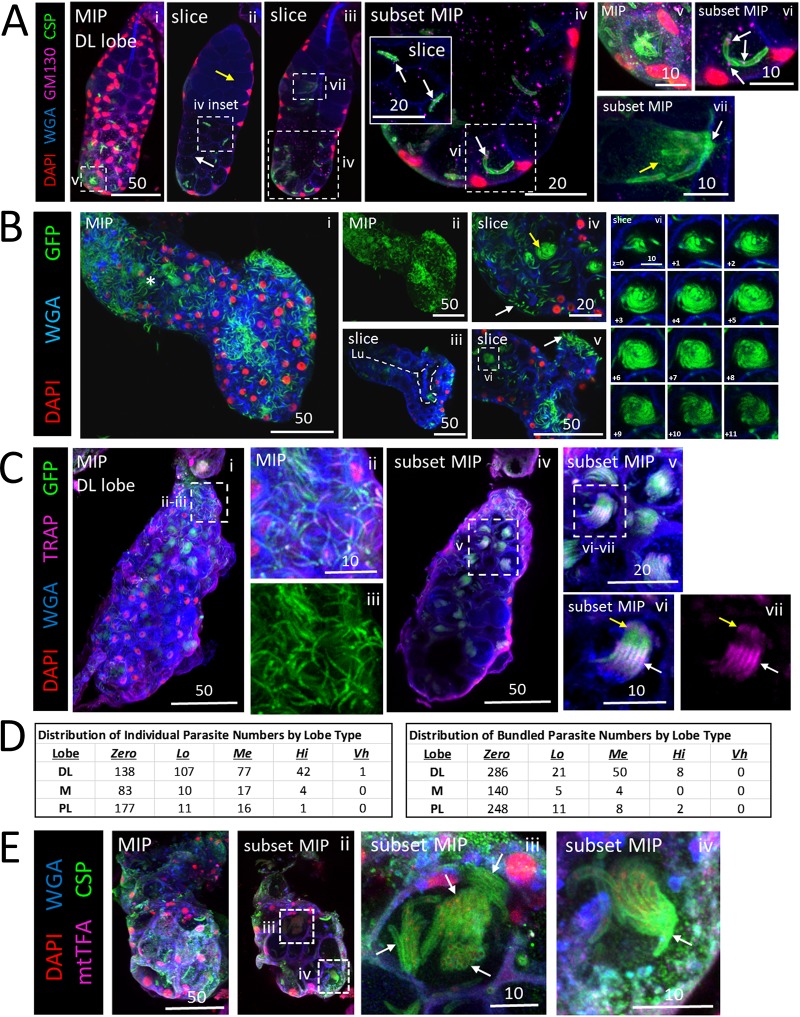
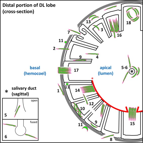
Plasmodium sporozoites (SPZs) must traverse the mosquito salivary glands (SGs) to reach a new vertebrate host and continue the malaria disease cycle. Although SGs can harbor thousands of sporozoites, only 10 to 100 are deposited into a host during probing. To determine how the SGs might function as a bottleneck in SPZ transmission, we have characterized Anopheles stephensi SGs infected with the rodent malaria parasite Plasmodium berghei using immunofluorescence confocal microscopy. Our analyses corroborate findings from previous electron microscopy studies and provide new insights into the invasion process. We identified sites of SPZ accumulation within SGs across a range of infection intensities. Although SPZs were most often seen in the distal lateral SG lobes, they were also observed in the medial and proximal lateral lobes. Most parasites were associated with either the basement membrane or secretory cavities. SPZs accumulated at physical barriers, including fused salivary ducts and extensions of the chitinous salivary duct wall into the distal lumen. SPZs were observed only rarely within salivary ducts. SPZs appeared to contact each other in many different quantities, not just in the previously described large bundles. Within parasite bundles, all of the SPZs were oriented in the same direction. We found that moderate levels of infection did not necessarily correlate with major SG disruptions or abundant SG cell death. Altogether, our findings suggest that SG architecture largely acts as a barrier to SPZ transmission.IMPORTANCE Malaria continues to have a devastating impact on human health. With growing resistance to insecticides and antimalarial drugs, as well as climate change predictions indicating expansion of vector territories, the impact of malaria is likely to increase. Additional insights regarding pathogen migration through vector mosquitoes are needed to develop novel methods to prevent transmission to new hosts. Pathogens, including the microbes that cause malaria, must invade the salivary glands (SGs) for transmission. Since SG traversal is required for parasite transmission, SGs are ideal targets for transmission-blocking strategies. The work presented here highlights the role that mosquito SG architecture plays in limiting parasite traversal, revealing how the SG transmission bottleneck is imposed. Further, our data provide unprecedented detail about SG-sporozoite interactions and gland-to-gland variation not provided in previous studies.

Keywords: malaria; mosquito; salivary gland; sporozoite.


Figures

FIG 1
FIG 1
Plasmodium sporozoites (SPZs) primarily invade the distal lateral lobes. Representative images of the entire depth (maximum intensity projection [MIP]) or partial depth (subset MIP) or single-slice confocal images of salivary glands (SGs) stained with DAPI (DNA, red), WGA (chitin/O-GlcNAcylation, blue), and either GFP (panels A and C to D; SPZs, green) or CSP (panel B; a SPZ protein, green) 18 to 24 days postinfection with P. berghei. Scale bar length units are micrometers. (A) Low and high magnification of SG with only the distal lobes infected and showing where SPZs were found: in secretory cell cytoplasms (panel ii, arrows), in secretory cavities (panel ii, arrowheads), in large, central, fluid-filled lumens (panel iii, arrows), in locations associated with the basement membrane (panel iv-vi, arrows), and rarely, inside the salivary duct (panel iv inset, arrow). The contrast in the inset in panel iv was uniformly enhanced to highlight the salivary duct SPZ and in panels v to vi to highlight the SPZs and secretory cell cytoplasms. The basement membrane (from the DIC channel; not shown) is marked by a dashed line (panels v and vi). (B) Representative images of PL (panel i) and M (panel ii) lobe infections. Multiple SPZs are observable (asterisks). (C) Number of lobes of different types imaged in this study (top) and number of SG lobes harboring parasites out of total infected SGs (bottom). (D) Total lobes imaged (left) and number of lobes imaged from uninfected SGs (right). (E) SG SPZ numbers differed from lobe to lobe, even within a single mosquito. A single SPZ was observed in one DL lobe (panel ii), in proximity to a fused salivary duct (panel iii). In another DL lobe from that mosquito, about 10 SPZs were oriented toward an irregular, round lumen (panel iv, arrow). Some infected lobe regions contained accumulations of what was likely shed CSP (panel iv, asterisk). (F) Representative image from a SG with secretory cell cytoplasms filled with SPZs (see the full lobe image in Fig. 4A and the description in the corresponding figure legend) that was used to determine that ∼40 sporozoites can occupy the cytoplasmic volume of a typical SG secretory cell (n = 25 cells). In this lobe, SPZs inside secretory cavities (slice image, asterisks) were easily discernible and were excluded from cytoplasmic SPZ counts. Two cells are outlined in white dashes. (G) Frequency and distribution of SPZs observed inside the salivary duct.
FIG 2
FIG 2
Quantification of salivary gland infection by Plasmodium sporozoites reveals invasion tendencies. (A) Examples of SGs with different numbers of invaded SPZs used for binning indicated as follows: no SPZs—Zero: 0 SPZ (panels i and ii); low numbers of SPZs—Lo: 1–10 SPZ (panels iii and iv); medium numbers of SPZs—Me: 11–100 SPZ (panels v and vi); high numbers of SPZs—Hi: 101–1000 SPZ (panels vii and viii); and very high numbers of SPZs—Vh: >1000 SPZ (panels ix and x). (B) Distribution of SPZ numbers in different lobes from infected SGs. (C) Localization of SPZs in the different lobes at different infection levels. At lower infection levels, most of the SPZs were associated with the basement membrane. At higher infection levels, most of the SPZs were found in secretory cavities. (D) Frequency of basal saliva accumulation by lobe in infected glands. (E) Frequency of basement membrane disruptions in different lobes from infected glands. (F) Frequency of lateral cytoplasmic disruptions in the different lobes at different infection levels. (G) Following a second, noninfective blood meal (BM) 23 days after P. berghei infection, SPZs were observed in similar SG locations: in secretory cell cytoplasms, in secretory cavities, and in the lumen (panels ii and iii, arrows). No increase in salivary duct occupancy was observed with greater SPZ numbers or with a second blood meal (panels G [iv] and C). Signal contrast was uniformly increased in panel G (iv) to highlight the salivary duct (SD). (H) In parallel, the infected mosquitoes in a second cage were given a second blood meal, and then female SGs were dissected and directly mounted on slides. Results showed individual (arrowheads) and bundled (arrows) sporozoites in similar numbers (quantified in panel I) and locations (secretory cell cytoplasm, secretory cavities, and lumen) and similar SG structural abnormality frequencies (quantified in panel J).
FIG 3
FIG 3
Salivary gland basement membrane and secretory cell traversal by sporozoites (SPZs) is associated with changes in the invasion and motility protein CSP. Representative 3D projection (MIP) or single-slice (slice) confocal microscopy images from salivary glands (SGs) stained with DAPI (DNA, red), either WGA (A, B, and D; chitin [O-GlcNAcylation], blue) or Nile red (C; lipids, blue), and either antisera against CSP (A, B, and D; SPZ protein, green) and mtTFA (A, B, and D; mitochondria, purple) or phalloidin (C; actin, green) 22 (A, B, and D) or 24 (C) days postinfection with P. berghei are shown. Scale bar length units are micrometers. (A) Distal lateral lobe showing large basement membrane disruptions (panel i) and rounded, likely dead parasites (panel ii). The examples shown represent cellular invasions and egressions by SPZs (panels iii to ix, asterisks). Three distinct CSP morphologies were observed: thick CSP SPZ coat (panel iv, arrow), thin CSP SPZ coat (panel v, arrow), and smaller tracks of shed CSP (panel iv, arrowhead). Thin CSP-coated SPZs are shown in secretory cavities (panel vi, asterisks). Thick CSP-coated parasites were observed invading secretory cells through the secretory cavities (panels vii and viii, asterisks) with a nearby basement membrane/cell disruption (panel viii, arrow). Rare SPZs with thick CSP coats were observed within damaged, dysmorphic SG secretory cell cytoplasms (panels ix and x, arrows). Perinuclear mtTFA enrichment in invaded cells is indicated (panel x, asterisks). (B) Low-magnification and high-magnification images of an infected distal lobe. A single SPZ (panel iii, white arrow) captured crossing the SG basement membrane (panels iii and iv, yellow arrow), a secretory cell cytoplasm (panels iii and iv, green arrow), a secretory cavity (panels iii and iv, red arrow), and lateral cytoplasmic extensions, which define the sides of the secretory cavity, of that cell and a neighboring cell (panels iii and iv, orange arrows) is shown. Contrast was uniformly enhanced in panel iv to highlight CSP and the SG cellular contents (WGA, mtTFA) associated with the invading sporozoite (panel v, white arrow) extending into the secretory cavity. Large basement membrane disruptions and large accumulations of shed CSP are shown (panels vi and vii, arrows). SPZs (panel viii, arrows) and CSP accumulations (panel viii, arrowheads) are observable. (C) Low-magnification image of a DL (panel i) with a SPZ (panels ii to iv) inside the lumen associated with a molecular halo (panels iii and iv, white arrow) that included Nile red and phalloidin staining accumulations. (D) Low-magnification image of SGs with only a few surrounding SPZs (panel i). Images of a single SPZ during invasion show that CSP staining was highly reduced beginning at the likely point of invasion (panels ii and iii, arrow). The basement membrane (purple dashed line) and plane of invasion (green dashed line) are indicated (panel iv).
FIG 4
FIG 4
Salivary gland architectural features associated with sporozoite (SPZ) accumulation. Images represent 3D projections over the entire salivary gland (SG) depth (MIP) or partial SG depth (subset MIP) or single-slice (slice) images from salivary glands stained with DAPI (DNA, red), WGA (chitin [O-GlcNAcylation], blue), and either GFP [A and C; SPZs, green] or CSP (B; SPZ protein, green) 23 (A and C) or 24 (B) days postinfection with P. berghei. Scale bar length units are micrometers. (A) SPZs near the basal surface of this DL lobe were individualized (panels i to iii), whereas SPZs in the apical region were tightly packed and oriented toward the salivary duct and lumen across much of the gland (panels iv and vii to ix). One secretory cavity containing SPZs was open to the lumen (panel iv, arrow). A single SPZ traversing a disruption in the salivary duct wall (panel v, vi, arrow) and a SPZ inside the salivary duct (panel ix, arrow) are shown. (B) Low-magnification image of infected glands (panel i). A fused duct terminus with sporozoites (SPZs) grouped in adjacent secretory cavities is shown in panels ii and iii. A DL lobe with a thick-walled salivary duct and swollen duct terminus connected to a small, mispositioned lumen with SPZs clustered at the duct wall and largely oriented toward the duct is shown in panel iv and in inset 1 in that panel. A possible passage, enriched for GFP, connecting the lumen to the adjacent secretory cavity is shown in panels iv (arrow) and iv (inset 2). SPZs were observed inside the lumen (panel iv, asterisk). (C) DL lobe with basement membrane and cell disruption (panels ii to iv, arrow) with two adjacent compartments filled with thick CSP-coated SPZs (panels ii to iv, arrowheads) in the same z focal plane (panel ii and iii) and in a different z focal plane (panel iv). More distally located secretory cells were open to the lumen (panel v), whereas more proximally located secretory cells had no periductal space and clustered SPZs near the duct (panel vi, arrows). (D) A corkscrewing SG with no lumen and a fused duct terminus (panel i) contained thick CSP-coated SPZs nearby (panel ii, arrow).
FIG 5
FIG 5
Apoptosis accompanying moderate invasion can be minimal, whereas large numbers of sporozoites (SPZs) can disrupt cell structure and saliva protein signal. (A and B) 3D projection (MIP) or single-slice confocal images of a representative distal lateral (DL) lobe stained with DAPI (nuclei, red), WGA (chitin [O-GlcNAcylation], blue), and antisera against GFP (SPZs, green) and either cleaved caspase 3 (A; CC3, purple) or the saliva protein Anopheles antiplatelet protein (B; AAPP, purple) 23 (A) or 24 (B) days postinfection with P. berghei. Scale bar length units are micrometers. (A) A distal lobe with large numbers of SPZs (panels ii, vi, and xi) had only two cells with accumulations of the apoptosis marker CC3 (panels ii, ix, and x, arrows) and only three small basement membrane disruptions (panels iii to v and vii to viii, asterisks). The images in panels ii to v and vi to viii are from two different focal planes. A neighboring DL lobe with a single CC3-positive cell is shown in panel vi (white arrow). Signal contrast was uniformly enhanced in panels ix to xi to highlight CC3 signal (panels ix and x) and SPZs (panel xi). (B) A DL lobe (panel i) with greatly different numbers of SPZs in the proximal and distal regions (panel ii). High SPZ numbers in the proximal region correlated with cell disruption (panel v, arrows [split secretory cell distal cytoplasms and lost basement membrane attachment]) and greatly reduced levels of AAPP saliva protein staining (panel v) compared to the distal region (panel vii).
FIG 6
FIG 6
Polarized organization of sporozoites (SPZs) within bundles. (A to D) 3D projection across entire SG depth (MIP) or part of the SG depth (subset MIP) images of a representative distal lateral (DL) lobe stained with DAPI (nuclear, red), WGA (chitin [O-GlcNAcylation], blue), and antisera against either GM130 (Golgi/cytoplasm, purple) and CSP (SPZ protein, green) (A), CSP alone (SPZ protein, green) (B), TRAP (SPZ protein, purple) and GFP (SPZs, green) (C), or mtTFA (SG cell cytoplasm, purple) and CSP (SPZ protein, green) (D) either 24 (A and B), 23 (C), or 22 (D) days postinfection with P. berghei. Scale bar length units are micrometers. (A) A DL lobe with only few SPZs in the proximal portion (panel ii, yellow arrow) and with more in the distal portion (panel ii, white arrow). Individual SPZs were observed (panel iv, inset) as well as SPZs interacting in a variety of quantities, including two (panels iv and vi), six or eight (panels iii and vii), or 10 to 12 (panels i and v). Note the concentrated shed CSP (panel vii, yellow arrow) at the salivary duct wall (panel vii, white arrow). GM130 was present in close proximity to the CSP coat of a SPZ in the secretory cavity (panels iv [and inset] and vi [white arrows]). (B) A bulbous DL lobe with high numbers of SPZs (panels I and ii) had an irregularly shaped lumen (panel iii, white dashed line). The asterisk in panel i marks the location of the salivary duct terminus. SPZs occupied the space between the basement membrane and the secretory cells (panels iv and v, white arrow), and SPZ groups were seen in a circling/swirling pattern (panels iv [yellow arrow] and vi [arrows]; see Movies S2 and S3). In panel vi, relative z positions are given in micrometers. (C) Infected DL lobe with both individualized SPZs (panels ii and iii) and SPZ bundles (panels iv to vii). TRAP localization (purple) to the apical tip (panels vi and vii, yellow arrows) and medial (panels vi and vii, white arrows) regions of SPZs (panels vi and vii) indicates that all SPZs within a bundle are similarly oriented anterior to posterior. (D) Frequencies of individual (left) or bundled (right) SPZs by lobe type. (E) Multiple SPZ bundles in a single secretory cavity (panel iii, arrows), with enrichment of CSP observable at basal end of a sporozoite bundle (panel iv, arrow), at a site of basement membrane disruption (panels i and ii, dashed boxes). CSP contrast was uniformly enhanced in panel iii to highlight the SPZs.
FIG 7
FIG 7
Roadblocks along the journey of Plasmodium sporozoites (SPZ) through Anopheles mosquito salivary glands. Schematic diagram depicting a cross-sectional view of a distal lateral (DL) lobe, infected by SPZs that encountered every barrier to salivary gland (SG) entry and exit observed in this study. Apical (SG lumen) and basal (mosquito hemocoel) compartments are noted (blue text). SPZs must interact with (step 1) and traverse (step 2) the basement membrane (gray), enter (step 2) and exit (step 3) the secretory cell cytoplasm (outlined in black) into the secretory cavity, enter the lumen (step 4), and finally enter the salivary duct (step 5) (the asterisk indicates the duct lumen) to proceed out of the mosquito during the next blood meal. Salivary ducts were sometimes (Fig. 1E, panel iii; see also Fig. 4B, panel ii, and D, panel ii) fused shut (step 6), as seen previously in male salivary glands (27, 28). Some lobes developed basement membrane and secretory cell disruptions (sections 7 and 8) that allowed SPZs a direct route (not requiring invasion) either to the lumen (step 7) or to the secretory cell cytoplasm (step 8). Some SPZs were seen in the secretory cell cytoplasm (sections 9 and 10), either as individuals (step 9) or groups (step 10). Rounded and/or fragmented SPZs (step 11) (36–38) were observed at the basement membrane and inside secretory cells. SPZs sometimes traversed secretory cell lateral cytoplasmic extensions en route to a neighboring secretory cavity (step 12). Other SPZs localized to pockets of lumenal saliva (step 13) were sometimes observed between the basement membrane and secretory cells (Fig. 1A, panel iv; see also Fig. 6B, panel ii). SPZs often bundled together when they reached a physical barrier, such as an aberrant WGA-positive chitinous wall (step 14) that sometimes lined the lumen (Fig. 5A). Some bundles cleared the barrier to enter the lumen (step 15), while in other cases, bundles represented a barrier that individual sporozoites navigated beyond (step 16). Some bundled SPZs traversed the basement membrane (step 17) (Fig. 6E, panel iv), exiting the SG and presumably entering the hemocoel. Certain SPZ groups, found primarily within secretory cavities, were captured by confocal imaging in a circling/swirling pattern (step 18) (Fig. 6B, panel vi; see also Movies S2 and S3).

Similar articles

Cited by

References

    1. World Health Organization. 2017. World malaria report 2017. World Health Organization, Geneva, Switzerland.
    1. Cibulskis RE, Alonso P, Aponte J, Aregawi M, Barrette A, Bergeron L, Fergus CA, Knox T, Lynch M, Patouillard E, Schwarte S, Stewart S, Williams R. 2016. Malaria: global progress 2000–2015 and future challenges. Infect Dis Poverty 5:61–61. doi:10.1186/s40249-016-0151-8. - DOI - PMC - PubMed
    1. Hemingway J, Shretta R, Wells TNC, Bell D, Djimdé AA, Achee N, Qi G. 2016. Tools and strategies for malaria control and elimination: what do we need to achieve a grand convergence in malaria? PLoS Biol 14:e1002380. doi:10.1371/journal.pbio.1002380. - DOI - PMC - PubMed
    1. Wang S, Dos-Santos ALA, Huang W, Liu KC, Oshaghi MA, Wei G, Agre P, Jacobs-Lorena M. 2017. Driving mosquito refractoriness to Plasmodium falciparum with engineered symbiotic bacteria. Science 357:1399. doi:10.1126/science.aan5478. - DOI - PMC - PubMed
    1. Pike A, Dong Y, Dizaji NB, Gacita A, Mongodin EF, Dimopoulos G. 2017. Changes in the microbiota cause genetically modified Anopheles to spread in a population. Science 357:1396. doi:10.1126/science.aak9691. - DOI - PubMed

Publication types