. 2019 Aug 6;10(4):e01834-19.
doi: 10.1128/mBio.01834-19.

Efficient Inhibition of Human Papillomavirus Infection by L2 Minor Capsid-Derived Lipopeptide

Affiliations

Efficient Inhibition of Human Papillomavirus Infection by L2 Minor Capsid-Derived Lipopeptide

Huan Yan et al. mBio. .

Abstract

The amino (N)-terminal region of human papillomavirus (HPV) minor capsid protein (L2) is a highly conserved region which is essential for establishing viral infection. Despite its importance in viral infectivity, the role of the HPV N-terminal domain has yet to be fully characterized. Using fine mapping analysis, we identified a 36-amino-acid (aa) peptide sequence of the L2 N terminus, termed L2N, that is critical for HPV infection. Ectopic expression of L2N with the transmembrane sequence on the target cell surface conferred resistance to HPV infection. Additionally, L2N peptide with chemical or enzymatic lipidation at the carboxyl (C) terminus efficiently abrogated HPV infection in target cells. Among the synthetic L2N lipopeptides, a stearoylated lipopeptide spanning aa 13 to 46 (13-46st) exhibited the most potent anti-HPV activity, with a half-maximal inhibitory concentration (IC50) of ∼200 pM. Furthermore, we demonstrated that the 13-46st lipopeptide inhibited HPV entry by blocking trans-Golgi network retrograde trafficking of virion particles, leading to rapid degradation. Fundamentally, the inhibitory effect of L2N lipopeptides appeared to be evolutionarily conserved, as they showed cross-type inhibition among various papillomaviruses. In conclusion, our findings provide new insights into the critical role of the L2N sequence in the HPV entry mechanism and identify the therapeutic potential of L2N lipopeptide as an effective anti-HPV agent.IMPORTANCE HPV is a human oncogenic virus that causes a major public health problem worldwide, which is responsible for approximately 5% of total human cancers and almost all cases of cervical cancers. HPV capsid consists of two structure proteins, the major capsid L1 protein and the minor capsid L2 protein. While L2 plays critical roles during the viral life cycle, the molecular mechanism in viral entry remains elusive. Here, we performed fine mapping of the L2 N-terminal region and defined a short 36-amino-acid peptide, called L2N, which is critical for HPV infection. Specifically, L2N peptide with carboxyl-terminal lipidation acted as a potent and cross-type HPV inhibitor. Taken together, data from our study highlight the essential role of the L2N sequence at the early step of HPV entry and suggests the L2N lipopeptide as a new strategy to broadly prevent HPV infection.

Keywords: L2N lipopeptide; entry inhibitor; human papillomavirus; minor capsid protein.


Figures

FIG 1
FIG 1
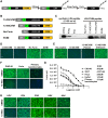
Fine mapping of the L2 N-terminal conserved region critical for HPV infectivity. (A) L2 similarity graph generated by using Vector NTI software, based on L2 sequences from 118 types of HPV (numbered according to HPV16 L2). The N-terminal region spanning aa 13 to 78 was selected for an alanine-scanning substitution assay. (B) L2 residue frequency graph generated by using WebLogo online software, based on aa 13 to 78 of L2 sequences from 118 types of HPV. The contribution of these residues to HPV infectivity (bottom) was summarized from HPV L2 fine mapping results in Fig. S2 and Table S1 in the supplemental material. (C) Illustration of 13 different L2 mutants (A18, A23, A28, A33, A38, A43, A48, A53, A58, A63, A68, A73, and A78) with sequential 5-amino-acid alanine substitutions. C20 and C28 were not changed in mutants A23 and A28. (D to F) CHO-K1 cells were infected with HPV-GFP/Lucia carrying L2 mutants. (D and E) GFP intensity (D) and Lucia activity (E) of infected cells were analyzed at 36 hpi. (F) L1 and L2 protein levels of input viruses were evaluated by using anti-HPV16 L1 (MD2H11) and anti-HPV16 L2 (2JGmab#5), respectively.
FIG 2
FIG 2
Ectopic expression of the L2 N-terminal region blocks HPV infection. (A) Schematic diagram of the Lucia-L2 N-terminal region fusion construct. F, furin cleavage sequence (RTKR); TM, transmembrane domain; C-ter, C-terminal region. (B) Illustration of the Lucia fusion constructs carrying different L2 N-terminal fragments and their anti-HPV activity. (+), >90% inhibition; (−), <10% inhibition; (+/−), 10 to 90% inhibition; No-Furin, R9K and R12K mutations in the furin cleavage sequence; No-SP, no secretion signal peptide. (C) FACS analysis of surface expression of Lucia-L2 N-terminal region fusion constructs in CHO-K1 cells with JWW-1 antibody. (D and E) HPV-GFP infection in CHO-K1 cells expressing the indicated Lucia-L2 N-terminal region fusion constructs. (D) Images were captured at 36 hpi. Bar, 200 μm. (E) Total GFP intensity was analyzed by using ImageJ.
FIG 3
FIG 3
Surface display of L2N aa 13 to 48 with the IL-2Rα transmembrane domain efficiently blocks HPV infection. (A) Schematic diagram of the secretion signal peptide (SP)-L2N-TM construct for surface display of L2 N-terminal aa 13 to 48. Tag, C-terminal tag. (B) Surface expression of the L2 N-terminal region on CHO-K1 cells stably expressing Lucia, Lucia-L2-6-55, or SP-L2N-TM. Surface expression levels were analyzed by flow cytometry with JWW-1 antibody. (C) Different cell lines expressing Lucia (mock) or SP-L2N-TM were infected by HPV-GFP. GFP intensity was analyzed at 36 hpi. Bars, 200 μm. (D) Expression levels of SP-L2N-TM mutants. (Top) Schematic diagram demonstrating the sequences of L2N aa 13 to 48 and highlighting the residues for mutagenesis analysis. (Bottom) Expression levels of SP-L2N-TM mutants in CHO-K1 cells at 3 days posttransduction were evaluated by immunoblotting with anti-FLAG antibody. (E) CHO-K1 cells expressing the indicated SP-L2N-TM mutants were infected with HPV-GFP. GFP intensity was analyzed at 36 hpi. Bar, 200 μm.
FIG 4
FIG 4
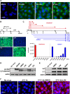
Inhibition of HPV infection by L2N lipopeptides. (A) Overview of L2N-CVIM peptide production. tSA, Twin-Strep-tag; F, furin cleavage site; CVIM, CAAX motif. (B) Schematic diagram of L2N-CVIM constructs. No-Furin, no furin cleavage sequence; SVIM, cysteine-to-serine mutation in the CVIM motif. (C) Quantification of L2N lipopeptides by JWW-1 immunoblotting with different amounts of a synthetic L2N peptide (13-55-no st). (D) HeLa cells were infected with HPV-GFP and treated with the indicated lipopeptides (∼500 nM) during inoculation. GFP intensity was analyzed at 36 hpi. Bar, 200 μm. (E) 13-48CVIM peptides were produced in 293T cells treated with 1 μM FTase inhibitor II (FtaseI) and 1 μM GGTase I inhibitor 2133 (GGtaseI) at 4 h posttransfection. The anti-HPV activity of these peptides was tested by an HPV-GFP infection assay in HeLa cells by coincubation with the inoculum for 16 h at a concentration of ∼500 nM. GFP intensity was analyzed at 36 hpi. Bar, 200 μm. (F) Inhibition of HPV-GFP infection by the 13-46st peptide (200 nM) in CHO-K1 cells, HeLa cells, and primary human epidermal keratinocytes. Bars, 200 μm. (G) IC50s of 13-46st peptide in different cells. Cells were infected with HPV-Lucia and treated with different concentrations of 13-46st during inoculation. Lucia activity was determined at 36 hpi (n = 2). IC50s were calculated by using GraphPad Prism software. (H) Cells were infected with GFP-carrying VSV, PIV3, HSV, or AAV at an MOI of 1 with or without treatment with 1 μM 13-46st during inoculation. Infection assays were conducted in CHO-K1 cells, except for HSV infection, which was conducted in Vero cells. GFP intensity was examined at 24 to 48 hpi. Bars, 100 μm.
FIG 5
FIG 5
The L2N lipopeptide blocks HPV entry without affecting HPV capsid priming and internalization. (A) Cell binding and uptake of L2N lipopeptide. HeLa cells were incubated with 500 nM FITC-m13-46st peptide for indicated times. Images were captured after three PBS washes. Bar, 25 μm. (B) L2N lipopeptide inhibits HPV-GFP entry. HeLa cells were treated with 500 nM L2N lipopeptide (13-46st) before (pre), during (co), or after (post) HPV-GFP inoculation. Mock, no peptide treatment. GFP intensity was analyzed at 36 hpi. Bar, 200 μm. (C) Peptide addition/withdrawal assays were performed as indicated in the schematic diagram (top). CHO-K1 cells were incubated with HPV-Lucia at 4°C for 2 h. The 13-46st lipopeptide (500 nM) was subsequently added or withdrawn from the medium 1, 2, 4, 8, and 16 h after viral attachment. Lucia activity was analyzed at 40 hpi. (D) CHO-K1 cells were infected with HPV-L2-PSTCD-3×FLAG (MOI of 50) and treated with the indicated chemicals: 500 nM dRVRK, 500 nM L2N lipopeptide 13-46st (L2N), 100 nM BafA1, and 1 μM γ-secretase inhibitor XXI. L2 immunoblotting was performed with anti-FLAG antibody at 16 hpi. (E) CHO-K1 cells were infected by HPV-GFP (MOI of 50) and treated with 500 nM L2N lipopeptide 13-46st, 100 nM BafA1, or both for 2 h. Intracellular L1 and L2 levels were determined by immunoblotting with MD2H11 and JWW-1, respectively. (F) HeLa cells were incubated with HPV-GFP (MOI of 50) at 4°C for 1 h and then shifted to 37°C for 4 h with or without 500 nM FITC-m13-46st. HPV L2 JWW-1 epitope exposure was analyzed by immunostaining with JWW-1 antibody (in red). Bar, 10 μm.
FIG 6
FIG 6
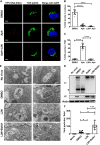
The L2N lipopeptide blocks TGN trafficking of HPV, leading to rapid virion degradation. (A) HPV retrograde trafficking to the TGN is blocked by L2N lipopeptide. HeLa cells were infected with EdU-labeled HPV (MOI of 50) and treated with DMSO, 3 μM aphidicolin (Aph), or 3 μM aphidicolin plus 500 nM 13-46st peptide (Aph+L2N). Cells were washed at 6 hpi and cultured for an additional 24 h with the indicated chemicals. HPV vDNA was visualized by a click reaction via Alexa 555 azide (red). Localization of the TGN (green) was analyzed by immunostaining with anti-P230 antibody. Bars, 5 μm. (B and C) Cellular distribution of EdU puncta. The ratios of EdU puncta in the nucleus (B) or in the TGN (C) were quantified based on representative images under each condition. P values were determined by ordinary one-way ANOVA with Dunnett’s multiple-comparison test (n = 10) ****, P < 0.0001. (D) CHO-K1 cells were infected with HPV-GFP (MOI of 50) and treated with 500 nM L2N lipopeptide 13-46st, 100 nM BafA1, or both for 24 h. Intracellular L1 and L2 levels were determined by immunoblotting with MD2H11 and JWW-1, respectively. *, full-length L1 (50 kDa); ●, 22-kDa L1 degradation fragment. (E and F) Ultrastructural analysis of intracellular HPV particles. (E) HeLa cells were infected with HPV at an MOI of 500 (about 10,000 particles/cell) for 16 h with or without treatment with 100 nM BafA1 and 500 nM 13-46st. Cells were processed for thin-section electron microscopy according to standard procedures, as described in Materials and Methods. Bars,100 nm. (F) Quantification of viral particles in representative images under each condition. One-way ANOVA (Kruskal-Wallis test) and Dunn’s multiple-comparison test were used for statistical analysis (n = 10). ***, P < 0.001; *, P < 0.05.
FIG 7
FIG 7
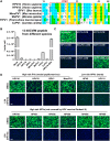
L2N peptide-mediated inhibition of various papillomavirus infections. (A) Sequence alignment of L2 N-terminal aa 13 to 48 (based on HPV16) from various papillomaviruses. (B and C) HeLa cells were infected with HPV16-GFP/Lucia and treated with L2N-13-48CVIM lipopeptides derived from various papillomaviruses (∼500 nM). The HPV16-2DK-CVIM (D31K and D43K) lipopeptide was included as a negative control. Lucia activity (B) and GFP intensity (C) were analyzed at 36 hpi (n = 2). Bar, 200 μm. (D) HeLa cells were infected with the indicated papillomaviruses (MOI of 1 to 5) and treated with or without 100 nM synthetic L2N lipopeptide of the HPV16 L2 sequence (13-46st). GFP intensity was examined at 36 hpi. Bars, 200 μm.

Similar articles

Cited by

References

    1. Tachezy R, Rector A, Havelkova M, Wollants E, Fiten P, Opdenakker G, Jenson B, Sundberg J, Van Ranst M. 2002. Avian papillomaviruses: the parrot Psittacus erithacus papillomavirus (PePV) genome has a unique organization of the early protein region and is phylogenetically related to the chaffinch papillomavirus. BMC Microbiol 2:19. doi:10.1186/1471-2180-2-19. - DOI - PMC - PubMed
    1. Garner MM, Hernandez-Divers SM, Raymond JT. 2004. Reptile neoplasia: a retrospective study of case submissions to a specialty diagnostic service. Vet Clin North Am Exot Anim Pract 7:653–671. doi:10.1016/j.cvex.2004.04.002. - DOI - PubMed
    1. de Villiers EM, Fauquet C, Broker TR, Bernard HU, zur Hausen H. 2004. Classification of papillomaviruses. Virology 324:17–27. doi:10.1016/j.virol.2004.03.033. - DOI - PubMed
    1. Lange CE, Favrot C, Ackermann M, Gull J, Vetsch E, Tobler K. 2011. Novel snake papillomavirus does not cluster with other non-mammalian papillomaviruses. Virol J 8:436. doi:10.1186/1743-422X-8-436. - DOI - PMC - PubMed
    1. Van Doorslaer K, Li Z, Xirasagar S, Maes P, Kaminsky D, Liou D, Sun Q, Kaur R, Huyen Y, McBride AA. 2017. The Papillomavirus Episteme: a major update to the papillomavirus sequence database. Nucleic Acids Res 45:D499–D506. doi:10.1093/nar/gkw879. - DOI - PMC - PubMed

Publication types

MeSH terms

LinkOut - more resources